| 图片: | |
|---|---|
| 名称: | |
| 描述: | |
- 谈东风病例8 Case T0008: 胃溃疡, 是癌吗?
| 姓 名: | ××× | 性别: | male | 年龄: | 71 |
| 标本名称: | |||||
| 简要病史: | History of renal cell carcinoma 12 years ago. GI bleeding for 4 months. | ||||
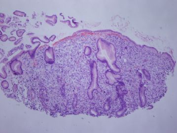
| 肉眼检查: | Endoscopically, an 1.8cm ulcer in the body of the stomach. Biopsy specimen. | ||||
This is an outside consultation case.
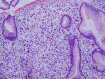
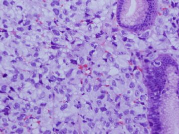
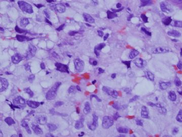
There are diffuse infiltrating epithelioid cells in the lamina propria. The cell borders are not clear. Some cells have clear cell changes (胞浆中有空泡), 间质内有少量的出血,结合病史要除外肾细胞癌.
Other common entities in the differential diagnosis include:
低分化腺癌(胃印戒细胞癌)
间叶源性肿瘤
恶黑
淋巴瘤
Histocytic lesions
Based on the morphological impression, a panel of immunostains was performed:
CK (focally, weakly +)
EMA (--)
Vimentin(focally +)
S-100(strongly ++)
CD45(--)
CD68(--).
Subsequently, HMB45 was performed:
HMB45 (++).
When I called to the clinician for the pathology results, he then stated that the patient also had a “mole” removed two years ago, but he did not have a pathology report of the “mole”.
Based on all the above information, it is most consistent with a metastatic melanoma (恶黑).
This is an example that 恶黑 can mimic many diseases. Whenever, you are not sure about an atypical lesion, 恶黑 should be always considered in the differential diagnosis.
-
本帖最后由 于 2010-05-30 06:20:00 编辑


