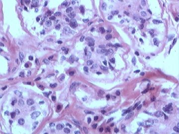
| 图片: | |
|---|---|
| 名称: | |
| 描述: | |
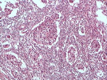
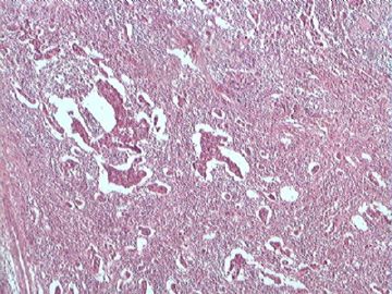
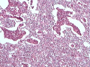
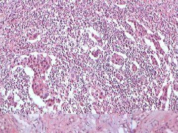
- B2679罕见乳腺癌类型(免疫组化结果公布)
| 姓 名: | ××× | 性别: | 女 | 年龄: | 33 |
| 标本名称: | 乳腺肿块 | ||||
| 简要病史: | 乳腺肿块3个月,无明显不适。体检有粘连,可活动。 | ||||
| 肉眼检查: | 肿块2X2厘米,切面灰白,质硬。 | ||||
-
本帖最后由 于 2010-05-24 08:17:00 编辑

- 许春雷
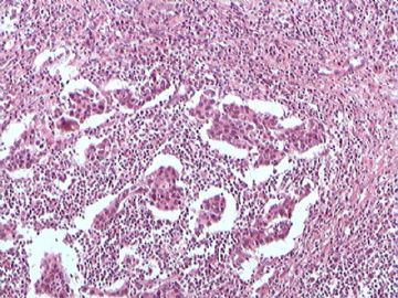
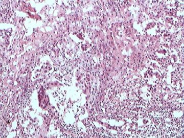
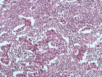
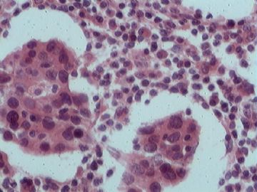
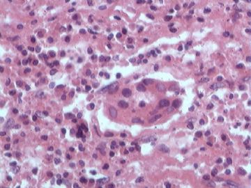
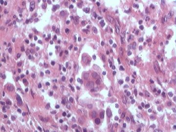
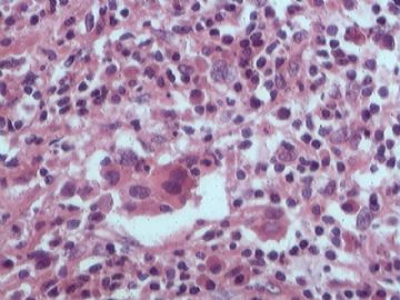
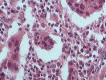
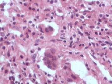
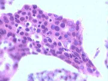
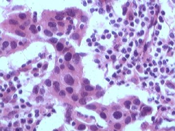
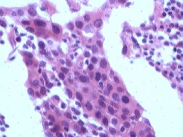
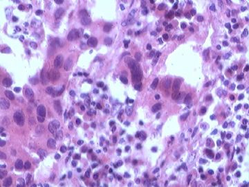
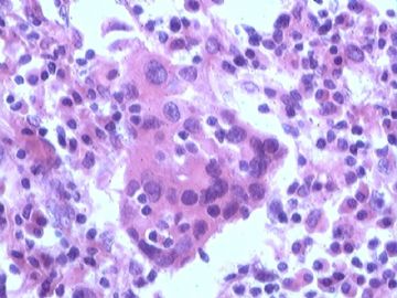
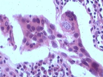
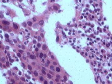
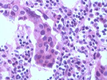
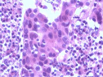
CK, ER positivity clearly demonstrates these large clusters are epithlial cells, but not histocytes. Possibally it is a ductal carcinoma with extensive inflammatory cell infilatrate.
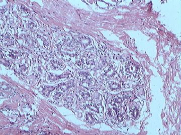
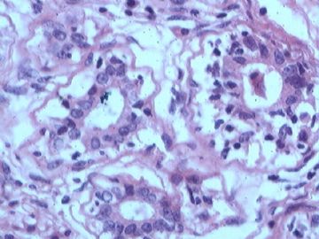
I am not very comfortable for one thing. For the H&E slides, almost all clusters seem isolated from the stroma tissue, floating amon the infalmmary cells. The H&E photos before the IHC photos show benign-looking ducts and lobules.
Should have more sections and try to find malignant tumor cells infilatrating the stroma
Seem that you are sure it is cancer. If you are sure it is not a lobular ca, you can call invasive ductal ca. In your comment you can mention the tumor mixed extensive acute and chronic inflammation.
many people think 炎性乳腺癌 is a clinical term if clinicians see tumor involving the skin. As pathologist we do not need to make this dx of 炎性乳腺癌

