
图1")
图2")
图3")
图4")
图5")
图6")
图7")
图8")
图9")
图10")
图11")
图12")
图13")
图14")
图15")
图16")
图17")
图18")


| 图片: | |
|---|---|
| 名称: | |
| 描述: | |
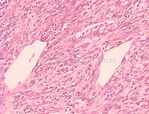
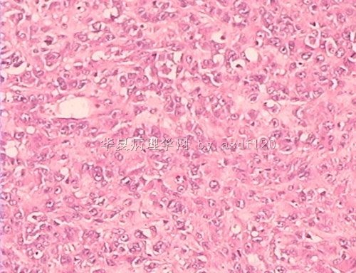
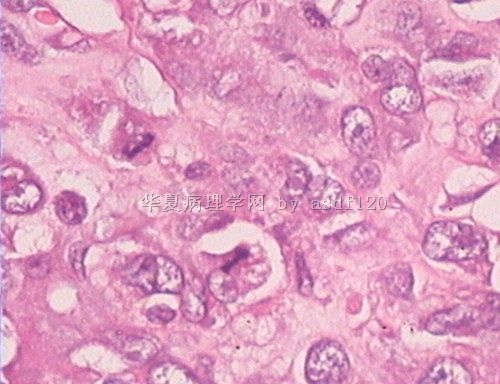
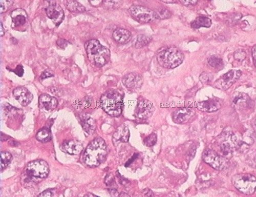
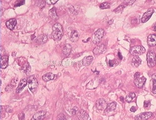
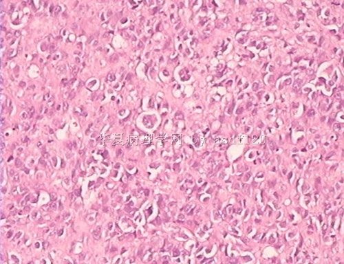
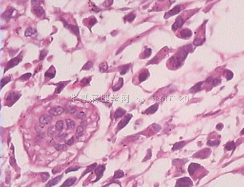
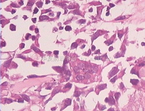
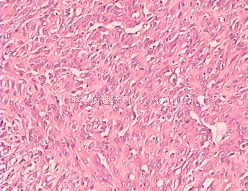
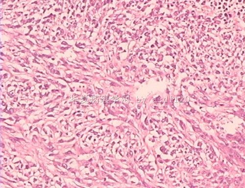
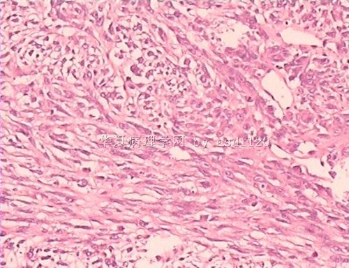
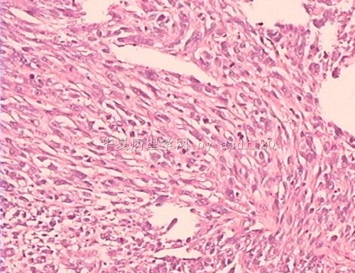
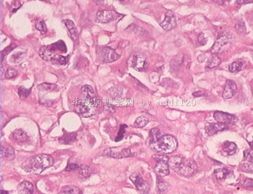
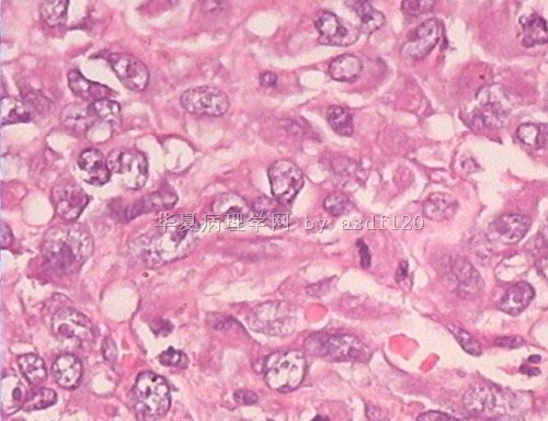
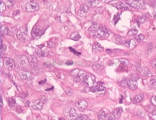
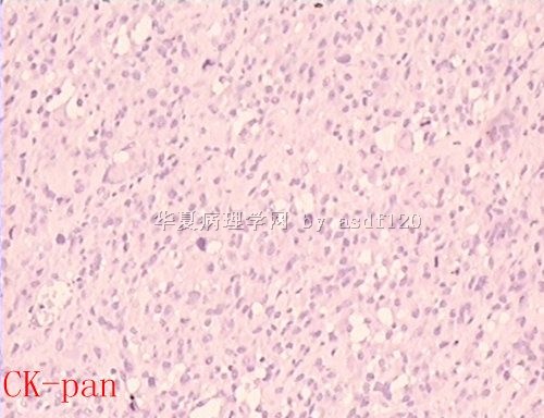
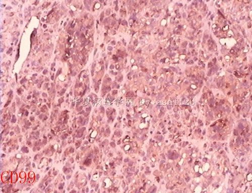
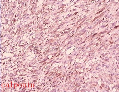
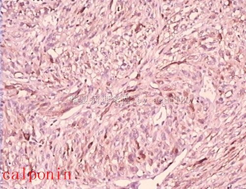
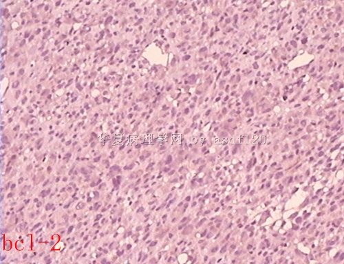
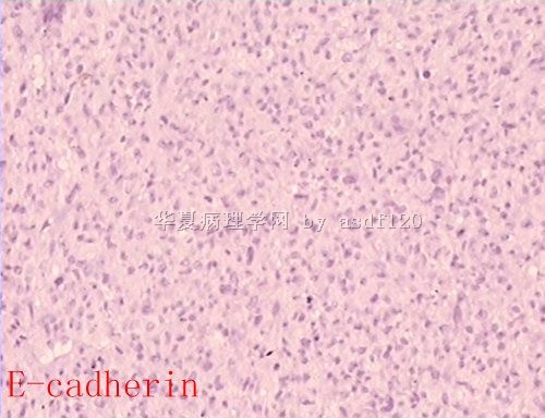
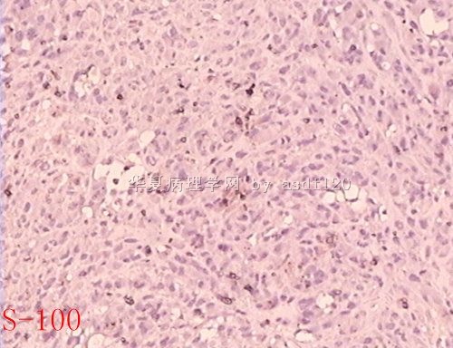
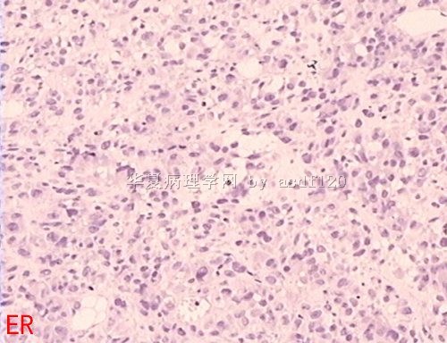
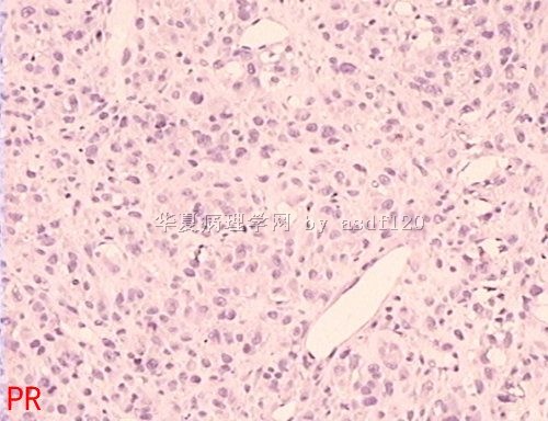
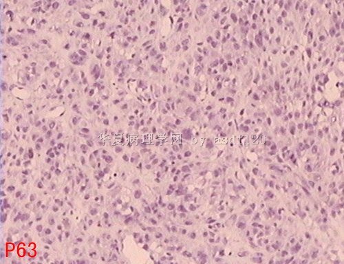
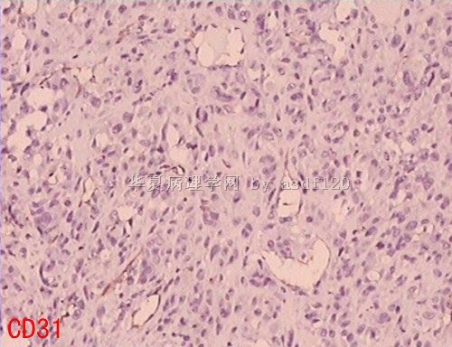
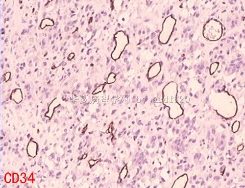
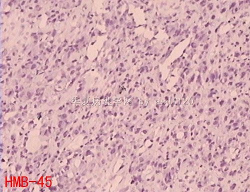
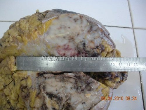
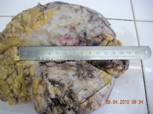
- B2663乳腺巨大肿瘤,请会诊!(附免疫组化)
1. The key for the case is carcinoma or sarcoma.
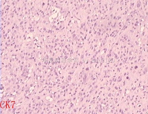
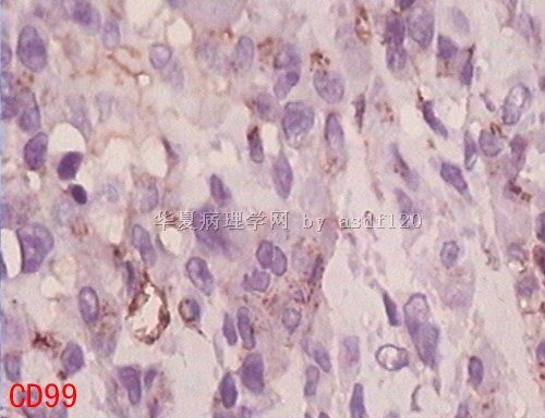
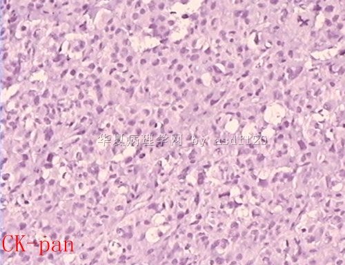
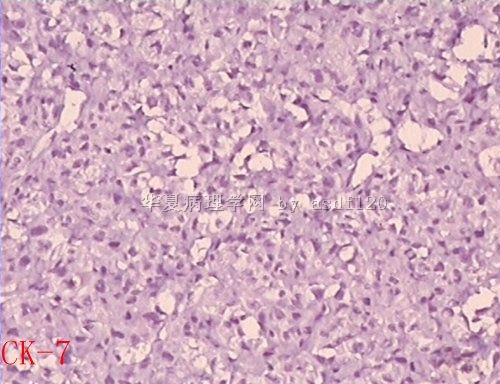
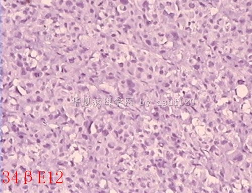
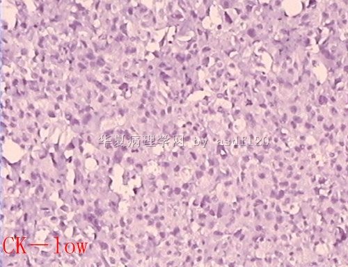
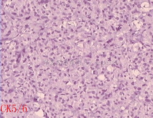
2. Notice that all stained epithelial markers (pan CK, EMA, CK7, CK19, CK-low, 34BE12, ck5/6) are negative. If enough sampling was enough, mostly it is not a carcinoma.
3. Then it is a sarcoma case. Next question is what type of sarcoma.
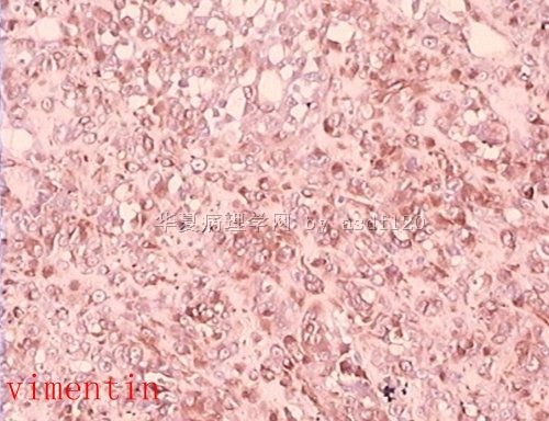
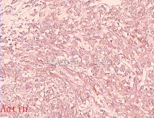
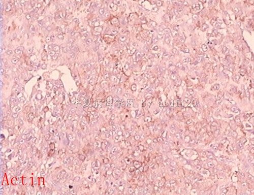
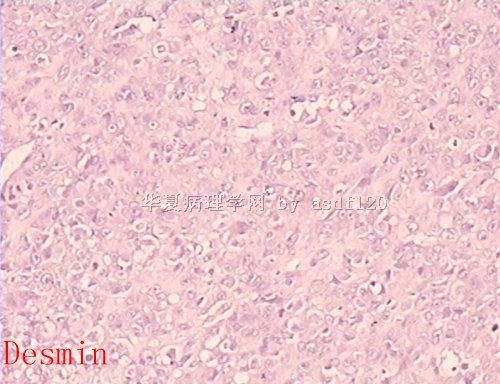
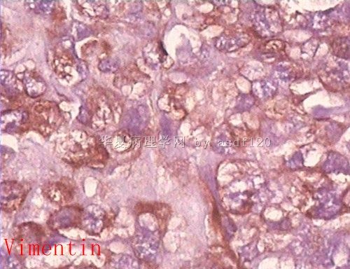
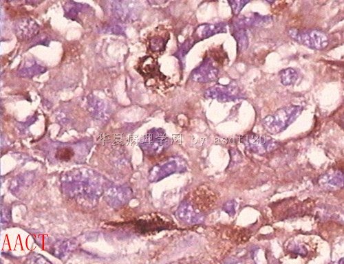
4. Calponin and actin are weakly positive (desmin negative). If they are true, it can be poorly differentiated leiomyosarcoma. What is AACT?
5. Do caldesmon stain
6. If smooth muscle origin cannot be determined, 多形性未分化肉类/恶性纤维组织细胞瘤is reasonal call.
| 以下是引用cqzhao在2010-4-14 11:14:00的发言:
1. The key for the case is carcinoma or sarcoma. 2. Notice that all stained epithelial markers (pan CK, EMA, CK7, CK19, CK-low, 34BE12, ck5/6) are negative. If enough sampling was enough, mostly it is not a carcinoma. 3. Then it is a sarcoma case. Next question is what type of sarcoma. 4. Calponin and actin are weakly positive (desmin negative). If they are true, it can be poorly differentiated leiomyosarcoma. What is AACT? 5. Do caldesmon stain 6. If smooth muscle origin cannot be determined, 多形性未分化肉类/恶性纤维组织细胞瘤is reasonal call.
thanks,α-1-Antichymotrypsin(AACT) |
-
本帖最后由 于 2010-04-18 22:16:00 编辑
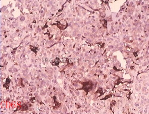
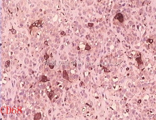
| 以下是引用zhuyan在2010-4-18 21:31:00的发言: 梭形肿瘤细胞Calponin ,Actin,均阳性,CD68为多核巨细胞阳性,似乎为反应性多核巨细胞,不太像恶性纤维组织细胞瘤中的肿瘤性多核巨细胞,我更倾向于诊断为平滑肌肉瘤-多形性。 |
1.Reasonal analysis.
2. In term of spindle cell sarcoma, stromal component of a malignant phyllodes is most common.
3. In term of pure sarcoma of breast, the most common one is angiosarcoma.
Seems above two are out of the game for this case.
4. Other types of sarcoma are very rare. Among them, leoomyosarcoma is relatively common.
5. Calponin and actin are at least weakly positive for this case even though desmin is negative. If possible, repeat these stains or add more smooth muscle marker stains.
I once saw more than 10 breast leiomyosarcoma, but never had a diagnosis of breast 恶性纤维组织细胞瘤.
6. in addition, smooth muscle marker stains are usually weak in high grade or poorly differentiated leiomyosarcomas.
7. In term of clinical treatment and prognosis, I do not think the pathologic diagnoses (types of sarcoma) will make big difference.


