| 图片: | |
|---|---|
| 名称: | |
| 描述: | |
- case1 宫颈小细胞癌; case2宫颈ADC; case 3 50 y/f月经增多
I showed a case here a few days ago. It was a very complicated law-suit case and I cannot find more photos now. So I deleted it because it is not good for education.Sorry for that.(几天前我贴了一个病例在这里。那是一个非常复杂的法律诉讼病例,由于我没有更多的图片,对于教学不是很好所以我删除了它。为此我深感抱歉。)
Our fellow showed an interesting case. I put here for your review.(我们的住院医有一个很有趣的病案,我贴在这里一起分享)
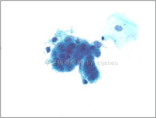
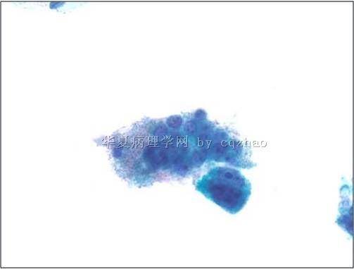
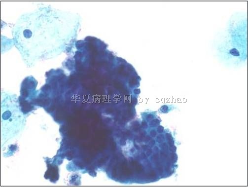
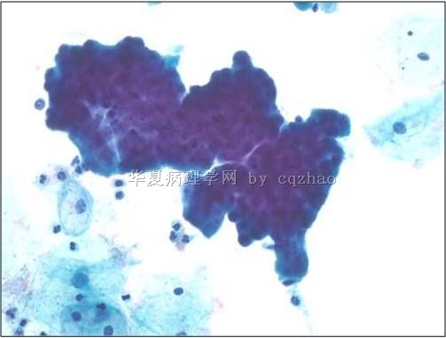
42 y women with LMP 5 days ago and no previous Pap history (女性,42岁,末次月经5天前,既往无巴氏检查)
-
本帖最后由 于 2010-05-07 07:28:00 编辑
-
yun_zhao123 离线
- 帖子:40
- 粉蓝豆:1
- 经验:42
- 注册时间:2010-05-06
- 加关注 | 发消息
-
yun_zhao123 离线
- 帖子:40
- 粉蓝豆:1
- 经验:42
- 注册时间:2010-05-06
- 加关注 | 发消息
“您是本帖的第 1696 位阅读者”哇哦,点击率真高啊!
这组图片涉及的应该是细胞群的诊断和鉴别诊断问题,赵老师又没有给我们可以对照的散在细胞,无疑更增加了诊断难度。
我想这样的细胞群首先要考虑的是腺和鳞的鉴别问题。看那么多人都考虑腺,我很没把握。但我认为就这几张图片来说,腺分化的证据不足,没有明显的三维、极化等结构特征,反而我看到部分水平排列细胞及隐约可见的核沟,这些都是鳞的特征。另外,即使是腺也不能肯定是宫内膜来源,如果说前面两个小细胞团还可以接受的话,宫内膜是不会出现后两个那么大的细胞团的。
其次看病变性质。细胞核浆比增大,染色质颗粒状,增粗不明显,大部分细胞有1-2个核仁。如果是鳞的话,肯定有问题,但如果是腺的话还不一定。
总之,这是个充满挑战与变数的病例,仅仅凭这几个细胞群诊断很没把握,如果一定要给个诊断的话,我会选择ASC-H/AGC,根据临床酌情处理。
It is not important how other people called the case. The importance is that how you will make your interpretation if you have the similar cases in your future practice.
Lession from this case:
We should be cautious when we see the cases with prominient and irregular 核仁 in endocervical cells.
-
本帖最后由 于 2010-04-26 20:17:00 编辑
3 years late this women had vaginal bleeding, the photos showed bx results. This is a very challenge case. You can determine or guess if this cluster of cells was the cancer cells. It may be yes, may be not.
Anyway, just hope every one knows that it is difficult for all pathologists to interpretate cervical glandular lesions in Pap cytology. This is one of the reasons why the rate of cervical adenocarcinoma continue to increase even though the rate of cervical carcinoma has declined internationally.
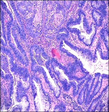
名称:图1
描述:图1
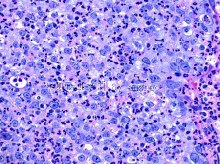
名称:图2
描述:图2


