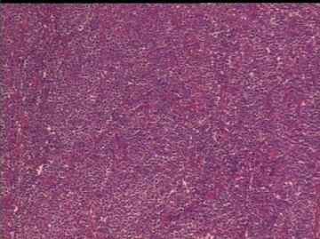
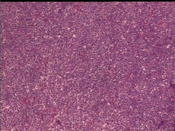
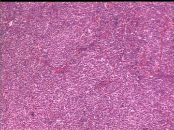
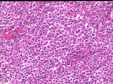
| 图片: | |
|---|---|
| 名称: | |
| 描述: | |
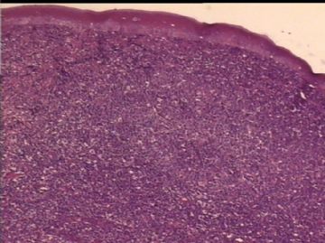
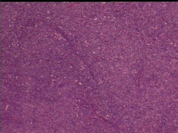
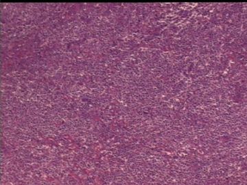
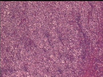
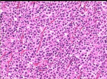
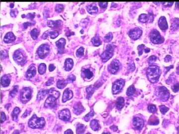
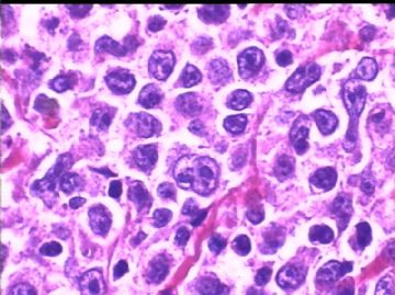
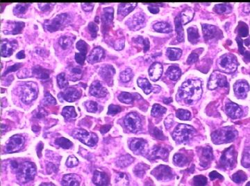
- 男/15岁 右侧扁桃体肿块, 淋巴瘤?IHC标记 2010-8-2
| 以下是引用wang4160在2010-5-27 14:41:00的发言:
发生在儿童的滤泡性淋巴瘤也不多!! 谢谢! |
WHO 2008 分类中已经列出了儿童滤泡性淋巴瘤的亚型
儿童滤泡性淋巴瘤(paediatric follicular lymphoma):肿瘤好发于青少年或年轻男性。病变局限,形态学上常有大滤泡,类似生发中心进行性转化(PTGC),CB常>15个/hpf,淋巴结的正常结构湮没。免疫组化显示CD10+、bcl6+、CD43+,但bcl2-。分子遗传学分析可证实为克隆性增生,但常无t(14;18)。肿瘤大多能完全治愈,通常不播散,预后好。

- 学无止境
-
panzenggang 离线
- 帖子:189
- 粉蓝豆:480
- 经验:246
- 注册时间:2008-01-09
- 加关注 | 发消息
| 以下是引用panzenggang在2010-8-3 8:14:00的发言:
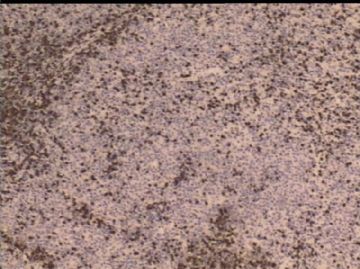
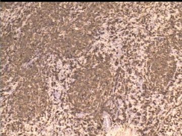
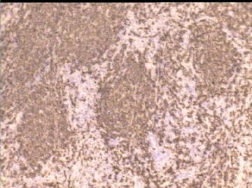
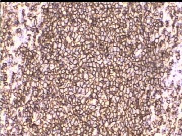
Dear Dr. Jin: Thanks a lot for your pictures. From these photos, I can clearly see a follicular proliferative pattern. But in picture 5, I couldn't tell if there is a positive staing of BCL2 in the follicular B-cells, and maybe there are a few cells with strong BCL2 staining, which may represent reactive T-cells in the germinal center. The last picture (Ki67), I see a relatively high Ki67 staining, but I also see a "negative zone" surrounding the follicle, is this a mantle zone? Also, usually low grade follicular lymphome will not have such a high Ki67 staining, but reactive germinal center can. If you have time, would you please explain to me? Thanks. I didn't see the original slides and it is hard for me to have a global picture of this case just based on the images, so I could be wrong. Sorry I can't type Chinese since I am in the office and we are not allowed to install programs ourselves. Have a nice day. |
非常感谢Dr.Pang对此病例的诊断意见。
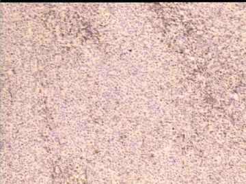
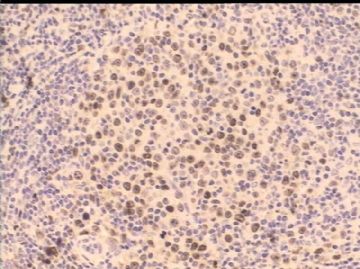
此例是会诊病例,原单位诊断为DLBCL。病变位于右侧扁桃体,肿块很大、抗炎治疗数月无效;左侧扁桃体正常。
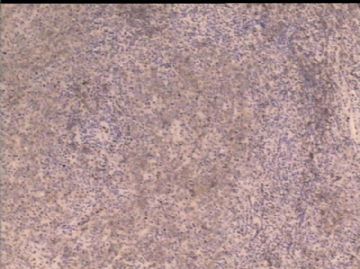
儿童滤泡型淋巴瘤是2008年WHO分类在滤泡型淋巴瘤中增加的新变型(Variant)。主要发生在颈部淋巴结、其他外周淋巴结和 Waldeyer Ring。 基本形态与成人型无区别,但有下列特点:1)病变比较局限、2)常常缺乏Bcl-2表达和t(14;18)、3)分级为3级、4)患者预后非常好,治愈率高。本病例 Bcl-2-、分级为3b。经RCHOP治疗6个疗程,肿瘤消失、全身情况良好。
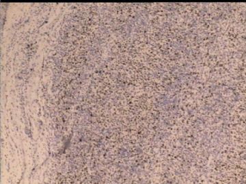
近期我会提供低倍镜图像,以利大家观察。谢谢!

- xljin8
-
panzenggang 离线
- 帖子:189
- 粉蓝豆:480
- 经验:246
- 注册时间:2008-01-09
- 加关注 | 发消息
Dear Dr. Jin:
Thanks a lot for your pictures.
From these photos, I can clearly see a follicular proliferative pattern. But in picture 5, I couldn't tell if there is a positive staing of BCL2 in the follicular B-cells, and maybe there are a few cells with strong BCL2 staining, which may represent reactive T-cells in the germinal center.
The last picture (Ki67), I see a relatively high Ki67 staining, but I also see a "negative zone" surrounding the follicle, is this a mantle zone? Also, usually low grade follicular lymphome will not have such a high Ki67 staining, but reactive germinal center can.
If you have time, would you please explain to me? Thanks. I didn't see the original slides and it is hard for me to have a global picture of this case just based on the images, so I could be wrong.
Sorry I can't type Chinese since I am in the office and we are not allowed to install programs ourselves.
Have a nice day.
-
panzenggang 离线
- 帖子:189
- 粉蓝豆:480
- 经验:246
- 注册时间:2008-01-09
- 加关注 | 发消息
-
本帖最后由 于 2010-08-02 05:08:00 编辑
I totally agree with "兰心".
We have to rule out a benign process in this 15 years old patient, particulary infectious mononucleosus. Need to do serology and clinical follow-up.
I see promient interfollicular expansion of the tonsil with a mixed population of cells, including immunoblasts, which is rather typical for viral infection.
Is there any IgH gene rearrangement or FISH done for this patient?
-
i_love_cells 离线
- 帖子:236
- 粉蓝豆:2467
- 经验:685
- 注册时间:2009-06-20
- 加关注 | 发消息

