| 图片: | |
|---|---|
| 名称: | |
| 描述: | |
- 肝脏病例第23例-地坛
先谢谢地坛医院的王主任提供好病例!
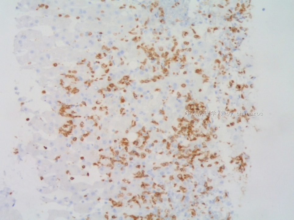
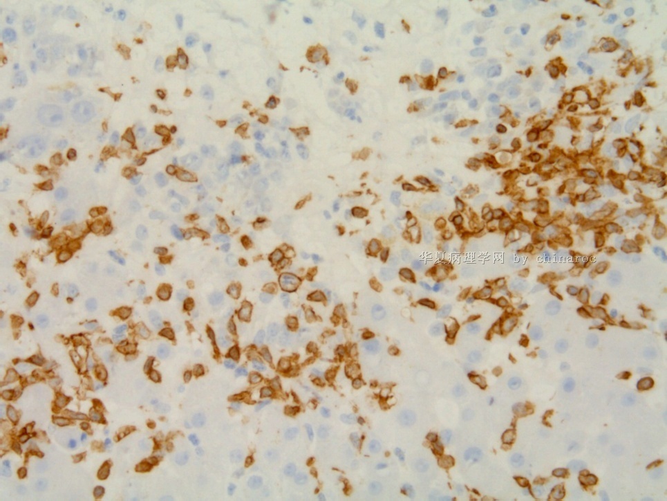
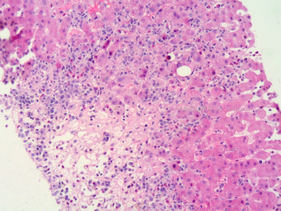
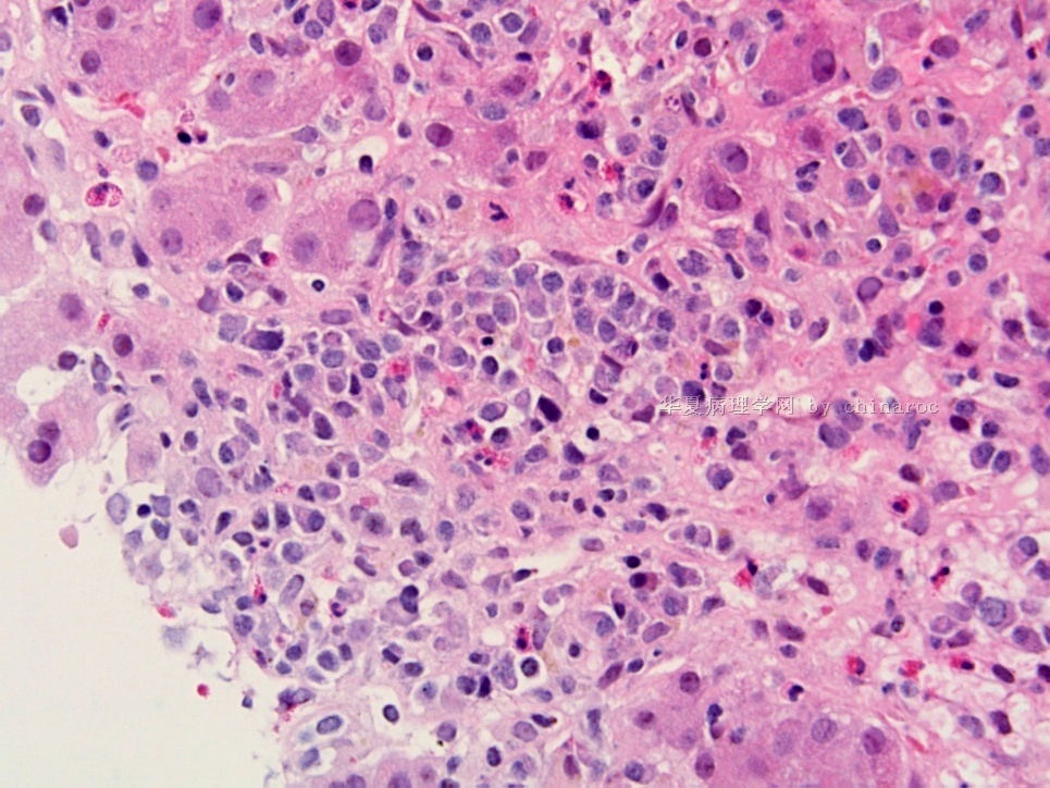
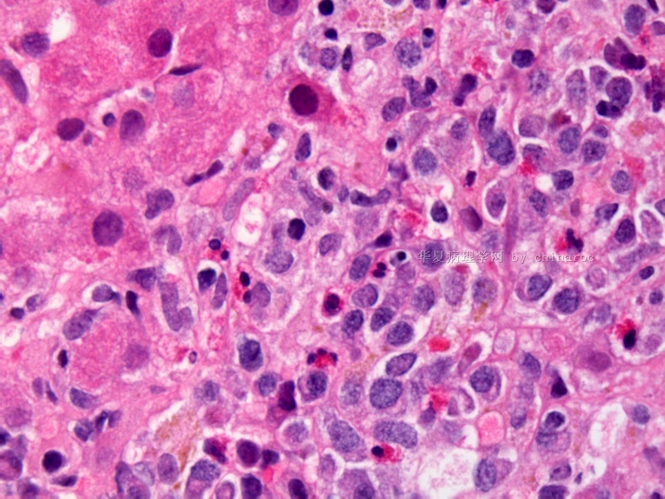
肝小叶不规则,中央静脉偏心,部分肝细胞浊肿、变性、增生,可见少量淤胆,(肝细胞内红色颗粒是代谢物沉积?看不明白)肝血窦扩张,汇管区浆细胞、嗜酸性粒细胞等炎细胞浸润,另有一些小异型细胞,胞浆灰红色,核偏位,核浆比高,可见核仁,浸润在肝细胞索间,部分在窦内。考虑:
1、淋巴造血系统疾病伴肝硬变:浆细胞性肿瘤?病人末梢血及骨髓涂片怎样?有无发热、肝、脾、淋巴结肿大等病史?
2、肝硬变伴肝细胞不典型增生?未见明确核内包涵体,不太支持,是不是药物或代谢性疾病引起?CK19、GCP-3,CD34。

- 广州金域病理
| 以下是引用天山望月在2010-2-14 21:54:00的发言:
肝小叶不规则,中央静脉偏心,部分肝细胞浊肿、变性、增生,可见少量淤胆,(肝细胞内红色颗粒是代谢物沉积?看不明白)肝血窦扩张,汇管区浆细胞、嗜酸性粒细胞等炎细胞浸润,另有一些小异型细胞,胞浆灰红色,核偏位,核浆比高,可见核仁,浸润在肝细胞索间,部分在窦内。考虑: 1、淋巴造血系统疾病伴肝硬变:浆细胞性肿瘤?病人末梢血及骨髓涂片怎样?有无发热、肝、脾、淋巴结肿大等病史? 2、肝硬变伴肝细胞不典型增生?未见明确核内包涵体,不太支持,是不是药物或代谢性疾病引起?CK19、GCP-3,CD34。 看得出天山望月的病理学功力深厚,这是一个比较有意思的病例,临床诊断为嗜酸性粒细胞增多症,一位肝脏病理学家诊断为药物性肝炎,而我却担心是淋巴系统的异常。 |

- 用心做事、真情做人!
-
Dr. Lin's opinion:
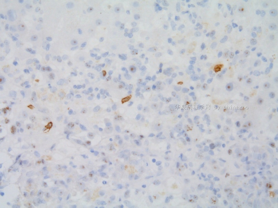
I have reviewed your case. There appear to be a mixed infiltrate of lymphoid cells, plasma cells, eosinophils and histiocytes. The CD138 may have not worked, it should have highlight the plasma cells. Because of the mixed infiltrate and the T cells are a mixed CD4 and CD8 cells, i think it is important to rule out infection, drug reaction and autoimmune disorder despite the fact that the pattern appears to be infiltrative. Some large cells may be residual hepatocytes. A marker for the hepatocytes will confirm that (the negative image of the lymphocyes may sometime better appreciated). In any event, I do not think that the disease in the liver can be explained by Hypereosinophilic syndrome because the predominant cell components in the liver is not eosinophils but lymphocytes.

- 用心做事、真情做人!
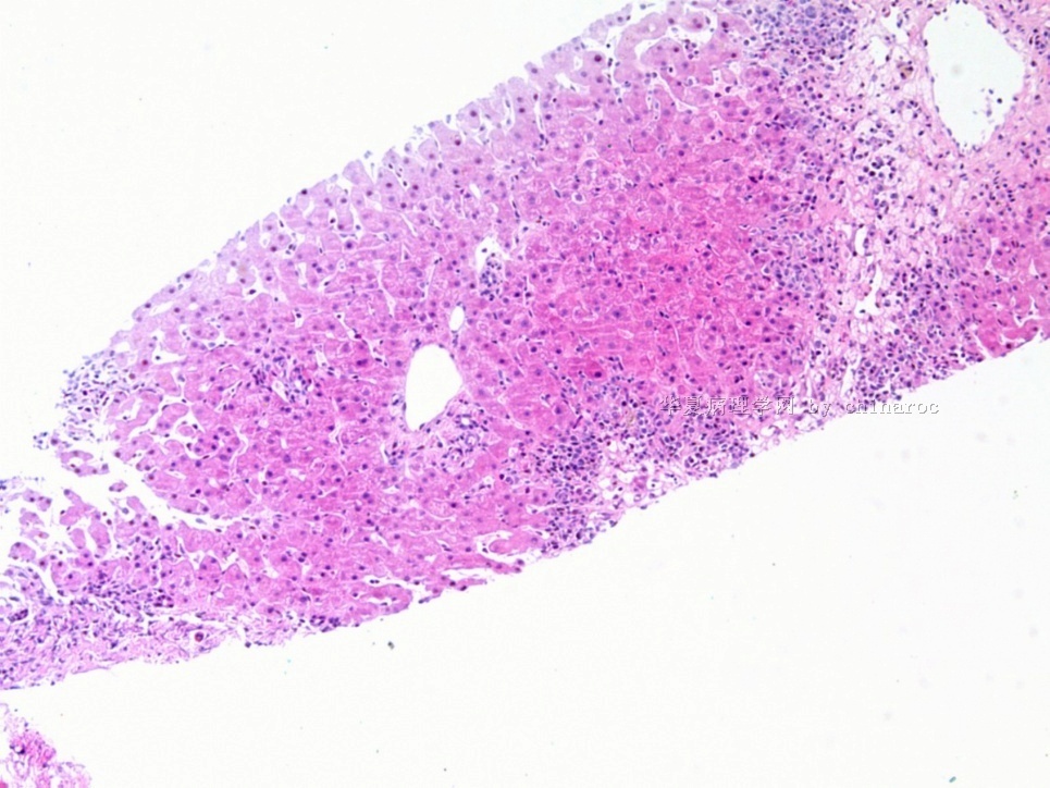
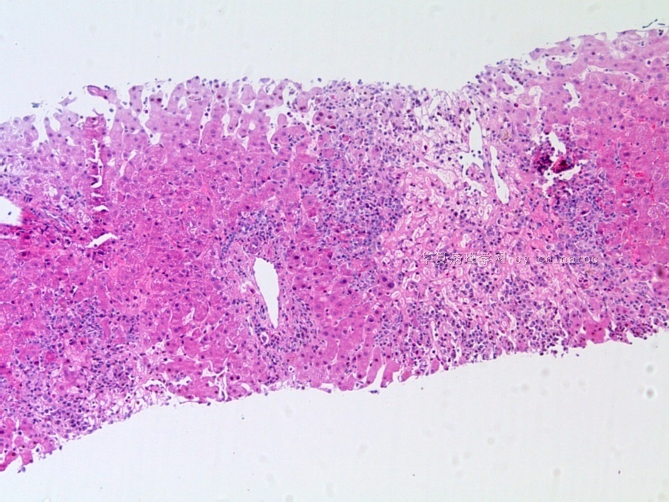
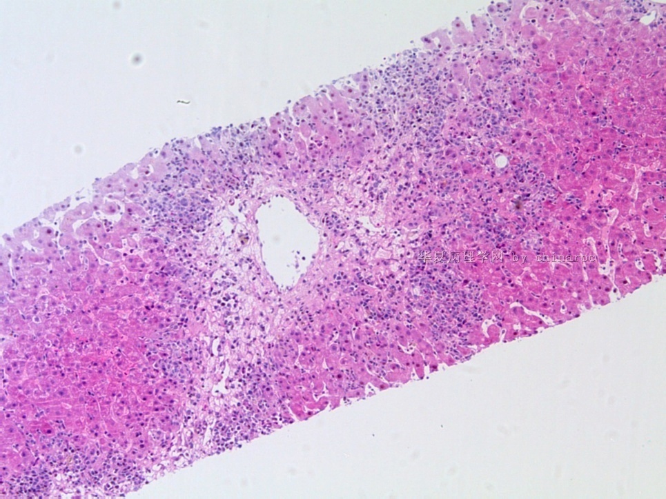
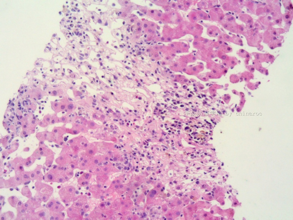


 ,最后病人怎样?大家有什么看法?请继续啊。