


| 图片: | |
|---|---|
| 名称: | |
| 描述: | |
- 20100123-十二指肠肿物
| 姓 名: | ××× | 性别: | 女 | 年龄: | 49岁 |
| 标本名称: | |||||
| 简要病史: | 发现右上腹包块4天, | ||||
| 肉眼检查: | 肿瘤位于十二指肠水平段,外生性生长,与胰腺肠系膜动脉关系密切,切除送检 | ||||

名称:图1
描述:图1

名称:图2
描述:图2

名称:图3
描述:图3

名称:图4
描述:图4

名称:图5
描述:图5

名称:图6
描述:图6

名称:图7
描述:图7

名称:图8
描述:图8

名称:图9
描述:图9

名称:图10
描述:图10

名称:图11
描述:图11
标签:
×参考诊断
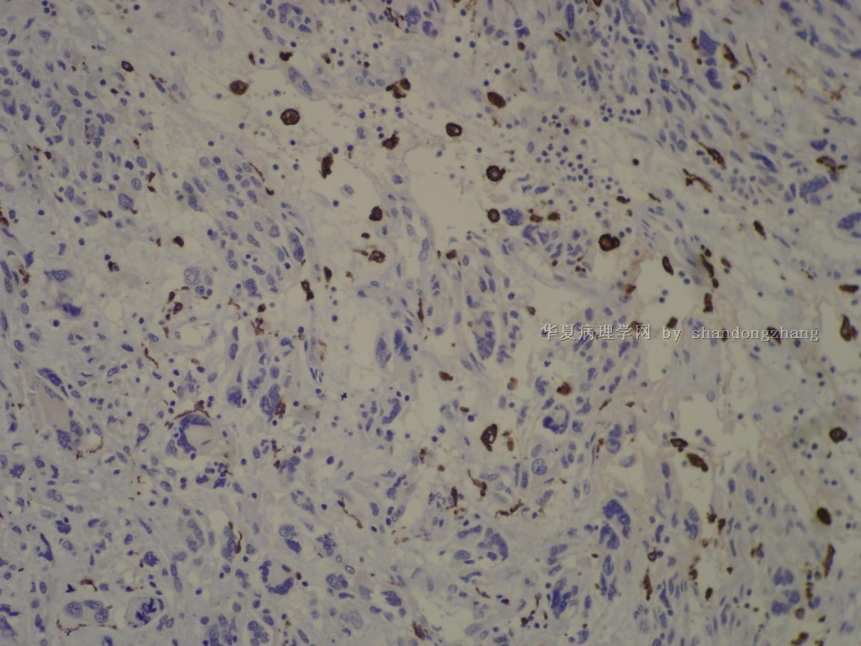
十二指肠胃肠间质瘤伴丰富的黏液样间质及含有“花环样”核的多核瘤细胞
关于粘液样基质的GIST参考文献:
Am J Surg Pathol. 1995 Jan;19(1):59-70.
Gastrointestinal stromal tumors with prominent myxoid matrix. Clinicopathologic, immunohistochemical, and ultrastructural study of nine cases of a distinctive morphologic variant of myogenic stromal tumor.
Arkadi M. Rywlin Department of Pathology & Laboratory Medicine, Mount Sinai Medical Center, Miami Beach, FL 33140.
Comment in:
Nine cases are presented of a distinctive morphologic variant of myogenic gastrointestinal stromal tumor characterized by an unusually prominent myxoid stromal background reminiscent of a neural neoplasm but lacking the immunohistochemical or ultrastructural features of peripheral nerve sheath or ganglionic differentiation. The patients included six women and three men aged 42 to 86 years (mean, 70). The lesions occurred in the stomach (seven cases) and small intestine (two cases) and ranged in size from 2.5 to 9.5 cm. They were described grossly as well circumscribed, unencapsulated, with a prominently myxoid and often cystic cut surface. Histologically, the lesions were composed of a proliferation of round, spindle, or stellate cells embedded in an abundant myxoid stroma. Histochemical stains showed strong positive reaction of the myxoid stromal background with alcian blue at pH 2.5; this staining reaction was abolished by treatment with hyaluronidase, indicating an abundance of connective tissue mucosubstances rich in hyaluronic acid. Immunohistochemical stains showed strong positivity of the tumor cells with vimentin antibodies in all cases and focal weak to moderate positive staining with muscle actin (HHF35) in eight cases and with desmin in two. Stains for keratin, S-100; epithelial membrane antigen, and collagen type IV were uniformly negative. Ultrastructural examination carried out in all cases showed features consistent with those previously described for myogenic gastrointestinal stromal tumors, namely, scattered mitochondria and prominent Golgi apparati, strands of rough endoplasmic reticulum, focal accumulation of intracytoplasmic microfilaments with occasional focal condensations, subplasmalemmal attachment plaques and immature cell junctions, focal extracellular basal lamina material, and surface-oriented micropinocytotic activity. The myxoid changes observed in these tumors may represent a secondary, nonspecific reaction pattern of the tumor cells to some noxious stimulus, or they may be a form of degenerative phenomenon such as that commonly observed in smooth-muscle tumors of the uterus and other sites. Myogenic gastrointestinal stromal tumors with prominent myxoid stroma should be distinguished from benign schwannoma of the stomach and gastrointestinal autonomic nerve tumors. Because of the differences in prognosis for these entities, immunohistochemical and ultrastructural examinations are recommended for the evaluation of gastrointestinal stromal neoplasms with prominent myxoid features.

- 王军臣
Arch Pathol Lab Med. 2004 Apr;128(4):440-3.
Malignant gastrointestinal leiomyosarcoma and gastrointestinal stromal tumor with prominent osteoclast-like giant cells.
Insabato L, Di Vizio D, Ciancia G, Pettinato G, Tornillo L, Terracciano L.
Dipartimento di Anatomia Patologica, Facolta di Medicina, Universita Federico II, Napoli, Italy. insabato@unina.it
CONTEXT: One case of leiomyosarcoma and one case of gastrointestinal stromal tumor with prominent osteoclast-like giant cells have so far been reported in the digestive tract. OBJECTIVE: To ascertain the clinicopathologic features and biologic behavior of these tumors, we report 3 additional cases of leiomyosarcoma of the gastrointestinal tract and one malignant gastrointestinal stromal tumor. DESIGN: Histologic and immunohistochemical examinations were performed. Clinical and follow-up data were recorded, and the literature was reviewed. RESULTS: The age of the patients ranged from 50 to 68 years (mean, 62 years). One of the lesions arose in the stomach, one in the ileum, and 2 in the colon. Three tumors showed a strong positivity for muscle actin and desmin and were diagnosed as leiomyosarcomas, 2 of them showing spindle cells and 1 of them showing epithelioid cells. The fourth tumor reacted strongly positive for c-Kit (CD117) and vimentin, and it was diagnosed as an epithelioid malignant gastrointestinal stromal tumor. All tumors were characterized by numerous osteoclast-like giant cells that were unevenly distributed and that, using immunohistochemistry, reacted strongly with CD68. CONCLUSIONS: Malignant stromal tumors with osteoclast-like giant cells of the gastrointestinal tract are rare entities, are more commonly of a myogenic origin such as leiomyosarcoma, and seem to have an aggressive behavior.
PMID: 15043462 [PubMed - indexed for MEDLINE]
- 王军臣
-
本帖最后由 于 2010-01-27 08:58:00 编辑
| 以下是引用海上明月在2010-1-26 23:12:00的发言:
同时伴有粘液样基质和破骨样巨细胞的恶性GIST的病例罕见,本例是一个非常好的罕见病例,值得学习借鉴。谢谢张老师! |
谢谢“海月”的关注及上传的资料
本例 花环样核的多核瘤细胞与文献上的破骨样多核巨细胞有些不同,CD163阴性。
Am J Surg Pathol 2005;29:52–68
Gastrointestinal Stromal Tumors of the Stomach A Clinicopathologic, Immunohistochemical, and Molecular Genetic Study of 1765 Cases With Long-term Follow-up 在这篇大宗1765例研究中描述了一系列的形态学改变:
Histologic Subtyping of Gastric
Stromal Tumors
Several histologic patterns occurred in sufficiently reproducible
constellations to allow for the delineation of eight histologic
subtypes, four representing spindle cell and four
epithelioid GISTs (Figs. 2, 3). A total of 1242 cases were
classified into these subtypes; 523 cases contained admixtures
of the ‘‘pure’’ subtypes or remained unclassified. Immunophenotypic
differences included common reduction of KIT positivity
in epithelioid tumors and less common smooth muscle
actin and desmin expression in sarcomatous tumors.
1. Sclerosing spindle cell GIST (n = 110): Paucicellular; extensive
extracellular collagen; slender spindle cells; no nuclear
atypia; low mitotic activity; common calcification (Fig. 2A,
B). Usually small incidental tumors, but 13% were .10 cm.
2. Palisading and vacuolated spindle cell GIST (n = 266):
Cellular; plump, uniform spindle cells; nuclear palisading;
perinuclear vacuolization; limited atypia and mitotic activity
rarely exceeding 10/50 HPFs (Fig. 2C, D).
3. Hypercellular, spindle cell GISTs (n = 139): Uniform,
densely packed, diffuse sheets of spindle cells; limited
atypia, nuclear palisading and perinuclear vacuolization
(Fig. 2E, F). Mitotic activity rarely exceeded 15/50 HPFs.
4. Sarcomatous spindle cell GISTs (n = 240): Spindled or oval
cells; diffuse atypia; tumor cells often in bundles separated
by myxoid stroma; mitotic activity over 20 per 50 HPFs;
nearly all .5 cm (Fig. 2G, H).
5. Sclerosing epithelioid GIST with a syncytial pattern (n =
270): Cohesive, uniform polygonal cells; indistinct cell
borders; diffuse collagenous matrix; multinucleation; low
mitotic rate (Fig. 3A, B).
6. Epithelioid GISTwith a dyscohesive pattern (n = 108): Large
polygonal cells with abundant cytoplasm; distinct cell borders;
discohesive; scant interstitial matrix; multinucleation;
possible focal atypia and low mitotic rate (Fig. 3C, D).
7. Hypercellular epithelioid GISTs (n = 78): Back-to-back epithelioid
cells; well-defined borders; nuclear atypia; higher
nucleocytoplasmic ratio than group 6; mitotic activity rarely
.10 per 50 HPFs (Fig. 3E).
8. Sarcomatous epithelioid GISTs (n = 31): Epithelioid to
rounded cells; well-defined borders; high nucleocytoplasmic
ratio; uniform nuclei, often prominent nucleoli; conspicuous
mitotic activity (.20/50 HPFs, Fig. 3F).
名称:图1
描述:图1
Arch Pathol Lab Med. 2002 Aug;126(8):972-4.
A malignant gastrointestinal stromal tumor with osteoclast-like giant cells.
Leung KM, Wong S, Chow TC, Lee KC.
Department of Pathology, Princess Margaret Hospital, Kowloon, Hong Kong.
Gastrointestinal stromal tumors (GISTs) are a heterogeneous group of mesenchymal tumors with a wide spectrum of histologic features and consistent expression of c-Kit. We describe an 85-year-old woman who presented with left lower quadrant abdominal pain and was subsequently diagnosed as having a malignant GIST. The tumor was composed of short fascicles of spindle cells. In addition to the presence of tumor giant cells, the tumor also demonstrated many osteoclast-like giant cells, a feature that has not been previously described in the literature. These giant cells expressed histiocytic markers CD68 and alpha(1)-antitrypsin but not c-Kit, a marker for GISTs. Electron microscopy showed no features of smooth muscle differentiation in the giant cells. The possible origin of the osteoclast-like giant cells is discussed in the context of immunohistochemical and ultrastructural characteristics.
- 王军臣


