
| 图片: | |
|---|---|
| 名称: | |
| 描述: | |
- 20100123-十二指肠肿物
| 姓 名: | ××× | 性别: | 女 | 年龄: | 49岁 |
| 标本名称: | |||||
| 简要病史: | 发现右上腹包块4天, | ||||
| 肉眼检查: | 肿瘤位于十二指肠水平段,外生性生长,与胰腺肠系膜动脉关系密切,切除送检 | ||||

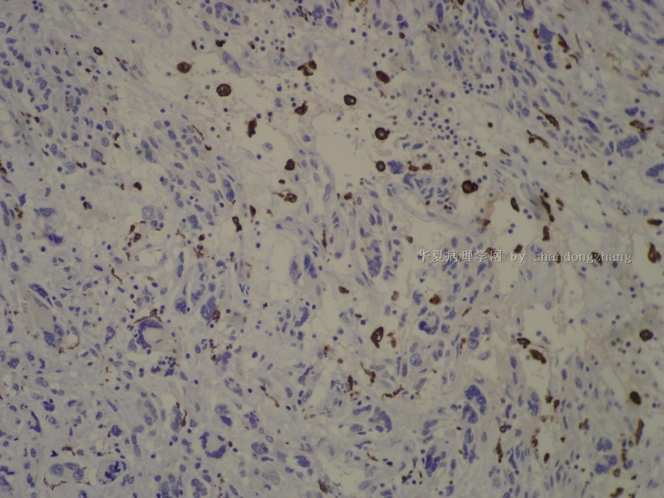
名称:图1
描述:图1

名称:图2
描述:图2

名称:图3
描述:图3

名称:图4
描述:图4

名称:图5
描述:图5

名称:图6
描述:图6

名称:图7
描述:图7

名称:图8
描述:图8

名称:图9
描述:图9

名称:图10
描述:图10

名称:图11
描述:图11
标签:
×参考诊断
十二指肠胃肠间质瘤伴丰富的黏液样间质及含有“花环样”核的多核瘤细胞
-
本帖最后由 于 2010-01-27 08:58:00 编辑
| 以下是引用海上明月在2010-1-26 23:12:00的发言:
同时伴有粘液样基质和破骨样巨细胞的恶性GIST的病例罕见,本例是一个非常好的罕见病例,值得学习借鉴。谢谢张老师! |
谢谢“海月”的关注及上传的资料
本例 花环样核的多核瘤细胞与文献上的破骨样多核巨细胞有些不同,CD163阴性。
Am J Surg Pathol 2005;29:52–68
Gastrointestinal Stromal Tumors of the Stomach A Clinicopathologic, Immunohistochemical, and Molecular Genetic Study of 1765 Cases With Long-term Follow-up 在这篇大宗1765例研究中描述了一系列的形态学改变:
Histologic Subtyping of Gastric
Stromal Tumors
Several histologic patterns occurred in sufficiently reproducible
constellations to allow for the delineation of eight histologic
subtypes, four representing spindle cell and four
epithelioid GISTs (Figs. 2, 3). A total of 1242 cases were
classified into these subtypes; 523 cases contained admixtures
of the ‘‘pure’’ subtypes or remained unclassified. Immunophenotypic
differences included common reduction of KIT positivity
in epithelioid tumors and less common smooth muscle
actin and desmin expression in sarcomatous tumors.
1. Sclerosing spindle cell GIST (n = 110): Paucicellular; extensive
extracellular collagen; slender spindle cells; no nuclear
atypia; low mitotic activity; common calcification (Fig. 2A,
B). Usually small incidental tumors, but 13% were .10 cm.
2. Palisading and vacuolated spindle cell GIST (n = 266):
Cellular; plump, uniform spindle cells; nuclear palisading;
perinuclear vacuolization; limited atypia and mitotic activity
rarely exceeding 10/50 HPFs (Fig. 2C, D).
3. Hypercellular, spindle cell GISTs (n = 139): Uniform,
densely packed, diffuse sheets of spindle cells; limited
atypia, nuclear palisading and perinuclear vacuolization
(Fig. 2E, F). Mitotic activity rarely exceeded 15/50 HPFs.
4. Sarcomatous spindle cell GISTs (n = 240): Spindled or oval
cells; diffuse atypia; tumor cells often in bundles separated
by myxoid stroma; mitotic activity over 20 per 50 HPFs;
nearly all .5 cm (Fig. 2G, H).
5. Sclerosing epithelioid GIST with a syncytial pattern (n =
270): Cohesive, uniform polygonal cells; indistinct cell
borders; diffuse collagenous matrix; multinucleation; low
mitotic rate (Fig. 3A, B).
6. Epithelioid GISTwith a dyscohesive pattern (n = 108): Large
polygonal cells with abundant cytoplasm; distinct cell borders;
discohesive; scant interstitial matrix; multinucleation;
possible focal atypia and low mitotic rate (Fig. 3C, D).
7. Hypercellular epithelioid GISTs (n = 78): Back-to-back epithelioid
cells; well-defined borders; nuclear atypia; higher
nucleocytoplasmic ratio than group 6; mitotic activity rarely
.10 per 50 HPFs (Fig. 3E).
8. Sarcomatous epithelioid GISTs (n = 31): Epithelioid to
rounded cells; well-defined borders; high nucleocytoplasmic
ratio; uniform nuclei, often prominent nucleoli; conspicuous
mitotic activity (.20/50 HPFs, Fig. 3F).
名称:图1
描述:图1


