共2页/32条首页上一页12下一页尾页
回复:32 阅读:4472
B2421乳癌术后胸水检测 (cqz-29)

cqzhao 离线

帖子:5518
粉蓝豆:1071
经验:5755
注册时间:2008-09-29
加关注  |  发消息
楼主 发表于 2009-12-31 11:14|举报|关注(1)
浏览排序[ 顺序 逆序 楼主 支持 精彩 ]  快捷回复
姓    名: ××× 性别:   年龄:  
标本名称:  
简要病史:  
肉眼检查:  

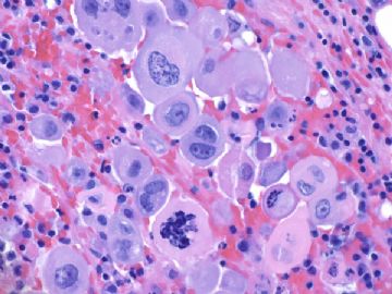
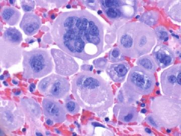
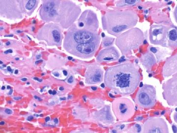
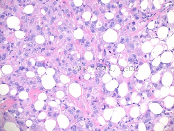
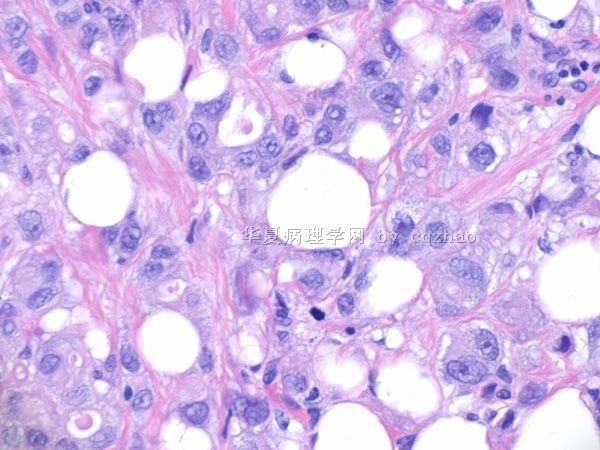
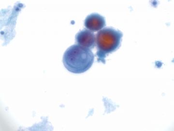
60 y/f with history of breast ca and had segmental mastectomy sereral years ago in other hospital. Patient present with pleural effusion.

F1. ThinPrep 400x

F2. cell block 400x

F3-4. cell block 600x


What will you do?

  • 乳癌术后胸水检测 (cqz-29)图1
    图1
  • 乳癌术后胸水检测 (cqz-29)图2
    图2
  • 乳癌术后胸水检测 (cqz-29)图3
    图3
  • 乳癌术后胸水检测 (cqz-29)图4
    图4
标签:乳腺多形性小叶癌
本帖最后由 于 2009-12-31 11:18:00 编辑
0

相关帖子

×参考诊断
(胸水)转移性乳腺多形性浸润性小叶癌,细胞学伴放化疗影响

笃行者 离线

帖子:2689
粉蓝豆:59
经验:3020
注册时间:2006-10-04
加关注  |  发消息
1 楼    发表于2010-03-24 19:10:00举报|引用
返回顶部 | 快捷回复
 赵老师的细胞学图片免疫组化做得如此漂亮!看这样的图片真是赏心悦目啊。
0
回复
signature
博学之,审问之,慎思之,明辨之,笃行之。

cqzhao 离线

帖子:5518
粉蓝豆:1071
经验:5755
注册时间:2008-09-29
加关注  |  发消息
2 楼    发表于2010-01-30 10:12:00举报|引用
返回顶部 | 快捷回复
本帖最后由 于 2010-01-30 10:15:00 编辑

First step:

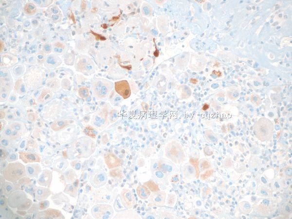
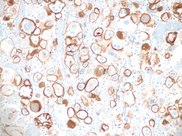
 I always do some stains for these kinds of fluid cytologic cases to distinguish mesothelial cells from epithelial cells first. 

Calretinin (mesothelial) and BerEP4 (epithelial)are the best two markers to choose.

名称:图1
描述:图1
名称:图2
描述:图2
0
回复

cqzhao 离线

帖子:5518
粉蓝豆:1071
经验:5755
注册时间:2008-09-29
加关注  |  发消息
3 楼    发表于2010-01-30 10:18:00举报|引用
返回顶部 | 快捷回复

 Above photo1 is calretinin and photo 2 is BerEP4.

Clearly they are epithelial cells. So it is a metastatic carcinoma.

The next step is to try to know where is the primary.

0
回复

cqzhao 离线

帖子:5518
粉蓝豆:1071
经验:5755
注册时间:2008-09-29
加关注  |  发消息
4 楼    发表于2010-01-30 10:21:00举报|引用
返回顶部 | 快捷回复
本帖最后由 于 2010-01-30 10:41:00 编辑

 CK7 and CK20 are useful markers for unknown primary.

CK7+/CK20-: breast, gynecological, lung.

CK-/CK20+: GI (especially low GI)

TTF-1: relative specific for lung

ER: gyn and breast

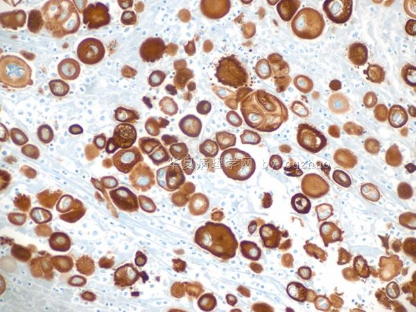
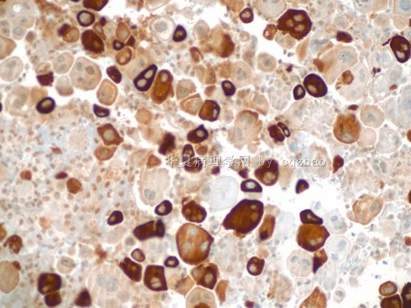
This patient had history of breast ca. I added mammaglobin.

F1. CK7

F2. CK20

F3. Mammaglobin

TTF-1 negative

ER positive

名称:图1
描述:图1

名称:图2
描述:图2
名称:图3
描述:图3
0
回复

cqzhao 离线

帖子:5518
粉蓝豆:1071
经验:5755
注册时间:2008-09-29
加关注  |  发消息
5 楼    发表于2010-01-30 10:27:00举报|引用
返回顶部 | 快捷回复
本帖最后由 于 2010-01-30 10:30:00 编辑

 Now we know most likely it was metastatic tumor from breast.

 Next question is what type of breast tumor.

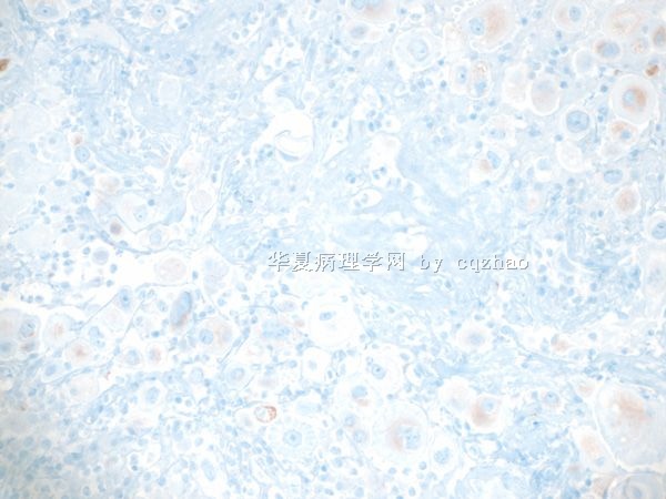
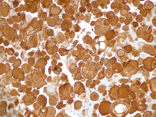
The cells in pleurAL fluid have some features of lobular carcinoma (photos in the top)

I stained e-cad and p-120.

F1. E-cadherin

F2. P-120

名称:图1
描述:图1
名称:图2
描述:图2
0
回复

cqzhao 离线

帖子:5518
粉蓝豆:1071
经验:5755
注册时间:2008-09-29
加关注  |  发消息
6 楼    发表于2010-01-30 10:32:00举报|引用
返回顶部 | 快捷回复
本帖最后由 于 2010-01-30 10:33:00 编辑

 Now I sure comfortable it is a metastatic lobular carcinoma.

I contacted the pathologist in the hospital where the patient had sgemental mastectomy and got the slides to review.


名称:图1
描述:图1

名称:图2
描述:图2
0
回复

cqzhao 离线

帖子:5518
粉蓝豆:1071
经验:5755
注册时间:2008-09-29
加关注  |  发消息
7 楼    发表于2010-01-30 10:40:00举报|引用
返回顶部 | 快捷回复
本帖最后由 于 2010-01-30 10:44:00 编辑

 Now I know that patient had breast pleomorphic lobular ca.

Also the patient recieved radiation chemotherapy.

The malignant cells in pleural effusion showed radiation and chemotherapy effect. This is why they are so ugly.

Wish more people like cytology.

Pathologists make diagosis based on the evidence, but not on the guess.

0
回复

bobol 离线

帖子:494
粉蓝豆:4572
经验:669
注册时间:2006-11-14
加关注  |  发消息
8 楼    发表于2010-01-13 12:36:00举报|引用
返回顶部 | 快捷回复

 转移性乳腺癌细胞?放疗相关(放射性肺炎)反应性间皮细胞?期待老师公布结果。

祝各位老师、前辈、同行新年快乐~

0
回复

jessica051604 离线

帖子:223
粉蓝豆:1
经验:898
注册时间:2010-01-18
加关注  |  发消息
9 楼    发表于2010-02-06 16:42:00举报|引用
返回顶部 | 快捷回复
 胸水中的核分裂不能作为诊断恶性肿瘤指标,多核在间皮细胞增生中也能见到. 学习了。
0
回复

cqzhao 离线

帖子:5518
粉蓝豆:1071
经验:5755
注册时间:2008-09-29
加关注  |  发消息
10 楼    发表于2009-12-31 11:21:00举报|引用
返回顶部 | 快捷回复
本帖最后由 于 2009-12-31 11:22:00 编辑

 This is my last case in 2009. Thank all people who read and/or attended discussion for these cases.

HAPPY NEW YEAR FOR ALL OF YOU. CZ

0
回复

nmchzhsh 离线

帖子:22
粉蓝豆:1
经验:22
注册时间:2010-03-08
加关注  |  发消息
11 楼    发表于2010-03-13 20:25:00举报|引用
返回顶部 | 快捷回复
 谢谢老师的讲解,见识了!
0
回复

笃行者 离线

帖子:2689
粉蓝豆:59
经验:3020
注册时间:2006-10-04
加关注  |  发消息
12 楼    发表于2010-01-01 21:19:00举报|引用
返回顶部 | 快捷回复

 对细胞学不是太熟悉,但感觉这些细胞核较大,核浆比增大,染色质较粗,核仁明显,有核分裂像,考虑是癌细胞。期待Dr.zhao解惑,谢谢!

HAPPY NEW YEAR TO YOU, Dr.Zhao.

0
回复
signature
博学之,审问之,慎思之,明辨之,笃行之。

cqzhao 离线

帖子:5518
粉蓝豆:1071
经验:5755
注册时间:2008-09-29
加关注  |  发消息
13 楼    发表于2010-01-01 21:20:00举报|引用
返回顶部 | 快捷回复

 自顶一下.

So beautiful photos. A few people are intersted. Feeling disappointed. ha....

0
回复

cqzhao 离线

帖子:5518
粉蓝豆:1071
经验:5755
注册时间:2008-09-29
加关注  |  发消息
14 楼    发表于2010-01-01 21:23:00举报|引用
返回顶部 | 快捷回复
以下是引用笃行者在2010-1-1 21:19:00的发言:

 对细胞学不是太熟悉,但感觉这些细胞核较大,核浆比增大,染色质较粗,核仁明显,有核分裂像,考虑是癌细胞。期待Dr.zhao解惑,谢谢!

HAPPY NEW YEAR TO YOU, Dr.Zhao.

We sent message here in the same time. You just did a few second before me.

Happy New year to you too. Thank you I learned the new word "笃" in 2009. cz
0
回复

文长江 离线

帖子:1042
粉蓝豆:139
经验:1438
注册时间:2007-03-21
加关注  |  发消息
15 楼    发表于2010-01-21 16:05:00举报|引用
返回顶部 | 快捷回复
 转移性乳腺癌细胞?
0
回复

xzy_nj 离线

帖子:14
粉蓝豆:1
经验:17
注册时间:2009-11-28
加关注  |  发消息
16 楼    发表于2010-01-21 19:05:00举报|引用
返回顶部 | 快捷回复
 腺癌细胞粘附性强一般很少散在分布,胸水中的核分裂不能作为诊断恶性肿瘤指标,多核在间皮细胞增生中也能见到.
0
回复
signature
认真对待一切

风雪夜行人 离线

帖子:251
粉蓝豆:27
经验:337
注册时间:2009-10-12
加关注  |  发消息
17 楼    发表于2010-01-24 15:19:00举报|引用
返回顶部 | 快捷回复
 期待IHC结果.
0
回复

wfbjwt 离线

帖子:6628
粉蓝豆:163
经验:7418
注册时间:2006-10-08
加关注  |  发消息
18 楼    发表于2010-01-24 15:50:00举报|引用
返回顶部 | 快捷回复
 漂亮的图片,谢谢分享!看到多核细胞我会想到伴破骨样细胞的乳腺癌,其次胞浆丰富我会想到恶黑。
0
回复
signature
嫁人就嫁灰太狼,学习要上华夏网。

zswx2008 离线

帖子:95
粉蓝豆:1
经验:658
注册时间:2008-05-25
加关注  |  发消息
19 楼    发表于2010-03-20 19:53:00举报|引用
返回顶部 | 快捷回复
  cqzhao 老师,你真棒!!!
0
回复
signature
让世界多一分绿色~

cqzhao 离线

帖子:5518
粉蓝豆:1071
经验:5755
注册时间:2008-09-29
加关注  |  发消息
32 楼    发表于2015-04-19 03:50:55举报|引用
返回顶部 | 快捷回复
引用 31 楼 n03553522119 在 2015-04-17 16:48:01 的发言:

 我是才上网不久的新生,看了很受益。


Glad you can learn some from the case

0
回复
回复:32 阅读:4472
共2页/32条首页上一页12下一页尾页
【免责声明】讨论内容仅作学术交流之用,不作为诊疗依据,由此而引起的法律问题作者及本站不承担任何责任。
快速回复
进入高级回复
您最多可输入10000个汉字,按 "Ctrl" + "Enter" 直接发送
搜索回复/乘电梯 ×
按内容
按会员
乘电梯
合作伙伴
友情链接