| 图片: | |
|---|---|
| 名称: | |
| 描述: | |
- B2421乳癌术后胸水检测 (cqz-29)
| 姓 名: | ××× | 性别: | 年龄: | ||
| 标本名称: | |||||
| 简要病史: | |||||
| 肉眼检查: | |||||
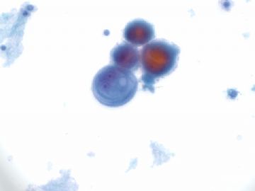
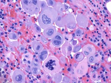
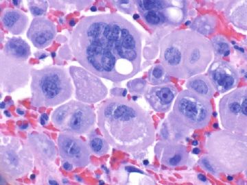
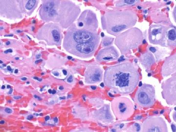
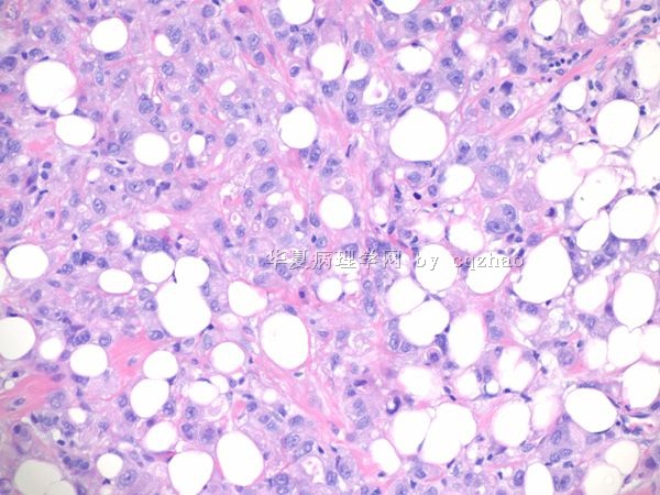
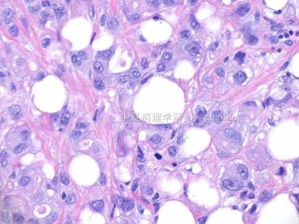
60 y/f with history of breast ca and had segmental mastectomy sereral years ago in other hospital. Patient present with pleural effusion.
F1. ThinPrep 400x
F2. cell block 400x
F3-4. cell block 600x
What will you do?
-
本帖最后由 于 2009-12-31 11:18:00 编辑
相关帖子
- • 右乳肿物诊断?
-
本帖最后由 于 2010-01-30 10:15:00 编辑
First step:
I always do some stains for these kinds of fluid cytologic cases to distinguish mesothelial cells from epithelial cells first.
Calretinin (mesothelial) and BerEP4 (epithelial)are the best two markers to choose.
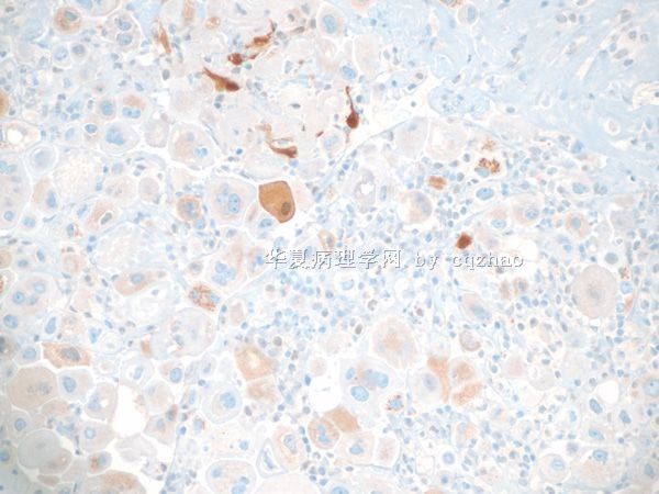
名称:图1
描述:图1
名称:图2
描述:图2
-
本帖最后由 于 2010-01-30 10:41:00 编辑
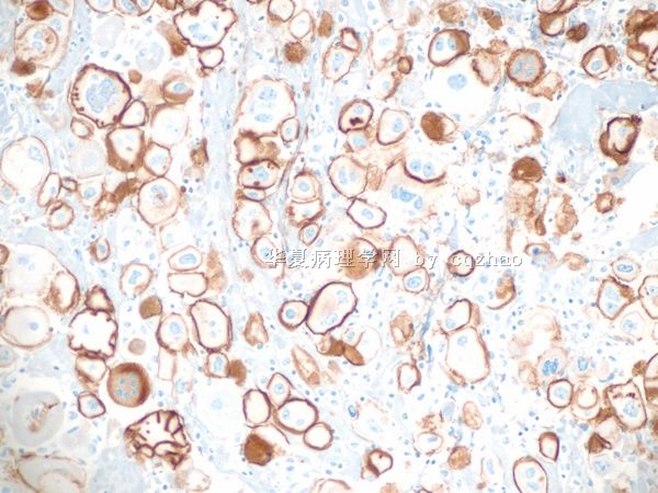
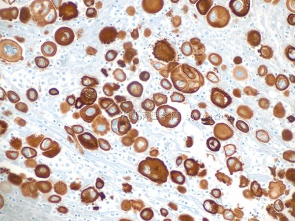
CK7 and CK20 are useful markers for unknown primary.
CK7+/CK20-: breast, gynecological, lung.
CK-/CK20+: GI (especially low GI)
TTF-1: relative specific for lung
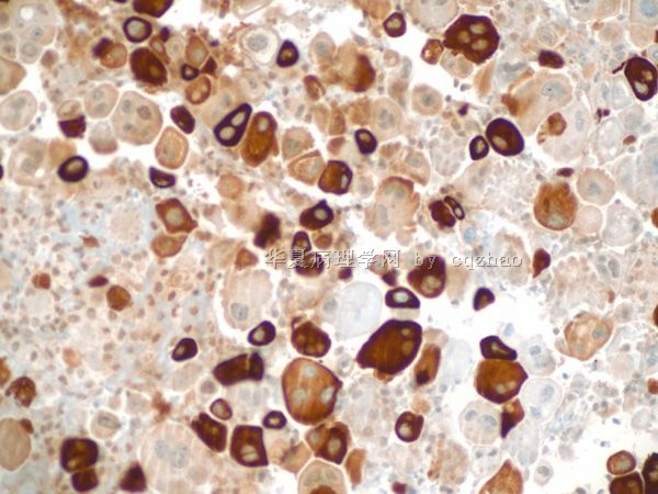
ER: gyn and breast
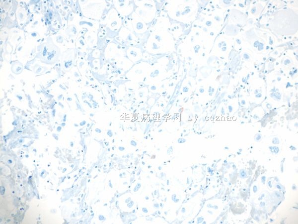
This patient had history of breast ca. I added mammaglobin.
F1. CK7
F2. CK20
F3. Mammaglobin
TTF-1 negative
ER positive
名称:图1
描述:图1
名称:图2
描述:图2
名称:图3
描述:图3
-
本帖最后由 于 2010-01-30 10:30:00 编辑
Now we know most likely it was metastatic tumor from breast.
Next question is what type of breast tumor.
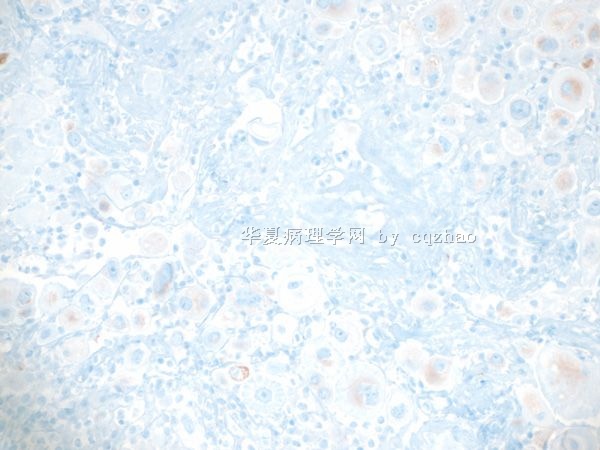
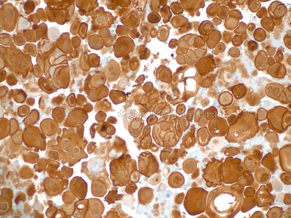
The cells in pleurAL fluid have some features of lobular carcinoma (photos in the top)
I stained e-cad and p-120.
F1. E-cadherin
F2. P-120
名称:图1
描述:图1
名称:图2
描述:图2
-
本帖最后由 于 2010-01-30 10:44:00 编辑
Now I know that patient had breast pleomorphic lobular ca.
Also the patient recieved radiation chemotherapy.
The malignant cells in pleural effusion showed radiation and chemotherapy effect. This is why they are so ugly.
Wish more people like cytology.
Pathologists make diagosis based on the evidence, but not on the guess.
| 以下是引用笃行者在2010-1-1 21:19:00的发言:
对细胞学不是太熟悉,但感觉这些细胞核较大,核浆比增大,染色质较粗,核仁明显,有核分裂像,考虑是癌细胞。期待Dr.zhao解惑,谢谢! HAPPY NEW YEAR TO YOU, Dr.Zhao. |
We sent message here in the same time. You just did a few second before me.
Happy New year to you too. Thank you I learned the new word "笃" in 2009. cz
