


| 图片: | |
|---|---|
| 名称: | |
| 描述: | |
- 神经病例1—地坛
-
本帖最后由 于 2009-12-31 12:54:00 编辑
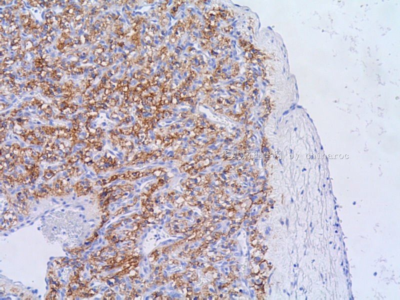
This looks like a WHO grade I hemangioblastoma. The anatomic location, rich vascularity (including capillaries and sinusoidal vessels) and neoplastic cells with plump, clear or vacuolated cytoplasm are very characteristic. The only differential diagnosis would be WHO grade I angiomatous meningioma, but I don't think it is. If you have any doubt, simply stain it for PR (positive for meningioma, negative for hemangioblastoma), EMA (positive for meningioma and negative for hemangioblastoma) and GFAP (some cells in hemangioblastoma are positive, and meningioma should be entirely negative) for confirmation.
象WHOI级血管母细胞瘤,解剖部位、丰富血管(包括毛细血管、血窦)以及胞浆丰富透亮空泡状的肿瘤细胞都很典型。唯一要鉴别的是WHOI级的血管瘤型脑膜瘤,我认为不是。如有疑问,可以染PR、EMA和GFAP证实:脑膜瘤PR+,EMA+,GFAP-;血管母细胞瘤PR-,EMA-,GFAP部分细胞阳。liguoxia71译

聞道有先後,術業有專攻
-
liguoxia71 离线

- 帖子:4174
- 粉蓝豆:3122
- 经验:4677
- 注册时间:2007-04-01
- 加关注 | 发消息
-
本帖最后由 于 2010-01-02 10:28:00 编辑
reticulin stain in hemangioblastoma: 围绕单个细胞或者小细胞团 surrounds individual cells or small clusters
关于EMA: hemangioblastoma 的细胞是阴性的, 但是文献报道偶尔有局部的阳性出现。
其实还有一个很重要的鉴别诊断是转移性的肾透明细胞癌RCC。而且很多病人有hemangioblastoma 的病人有Von Hippel Lindau syndrome所以也会有肾透明细胞癌, 文献上还有极个别的肾透明细胞癌转移到呵mangioblastoma的。
RCC与hemangionblastoma的鉴别诊断:
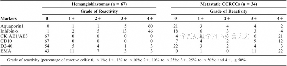
Am J Surg Pathol. 2008 Jul;32(7):1051-9.
Immunohistochemical markers to distinguish between hemangioblastoma and metastatic clear-cell renal cell carcinoma in the brain: utility of aquaporin1 combined with cytokeratin AE1/AE3 immunostaining.
Weinbreck N, Marie B, Bressenot A, Montagne K, Joud A, Baumann C, Klein O, Vignaud JM.
Department of Pathology, Nancy University Hospital, Nancy, France.
Distinguishing hemangioblastomas from metastatic clear-cell renal cell carcinomas (CCRCCs) in the brain is a diagnostic challenge owing to similar clinical and morphologic presentations. Inhibin-alpha and aquaporin1 were shown as positive markers of hemangioblastoma, but are not totally reliable distinguishing hemangioblastoma from metastatic CCRCC. This study shows that the diagnosis can be achieved using a combination of markers. To identify the panel of markers useful for this differential, 67 hemangioblastomas and 34 metastatic CCRCCs were analyzed using a panel of antibodies including aquaporin1, inhibin-alpha, D2-40, cytokeratin AE1/AE3, epithelial membrane antigen, and CD10. The study confirms the usefulness of aquaporin1 (97% sensitivity, 83% specificity) and inhibin-alpha (88% sensitivity, 79% specificity) as positive markers of hemangioblastoma and shows that aquaporin1 is a superior positive marker versus inhibin-alpha for the differential. Positivity of tumor cells with cytokeratin AE1/AE3 is the signature of a metastatic CCRCC (100% specificity, 88% sensitivity) and CD10 expression as well (100% specificity, 79% sensitivity). The combined use of aquaporin1 and AE1/AE3 yields a high degree of sensitivity and specificity to differentiate between hemangioblastoma and metastatic CCRCC. All tumors but one aquaporin1 positive and cytokeratin AE1/AE3 negative (65/66) correspond to hemangioblastomas (97% sensitivity, 97% specificity, 98.5% diagnostic positive predictive value). Tumors with the opposite profile, aquaporin1 negative, and cytokeratin AE1/AE3 positive, (25/25), correspond to metastatic CCRCC (74% sensitivity, 100% specificity, 100% diagnostic positive predictive value). In summary, aquaporin1 is the most sensitive positive marker of hemangioblastoma. Despite its moderate specificity, when used in combination with epithelial marker AE1/AE3, it allowed to reliably distinguish hemangioblastoma from metastatic CCRCC.
名称:图1
描述:图1
-
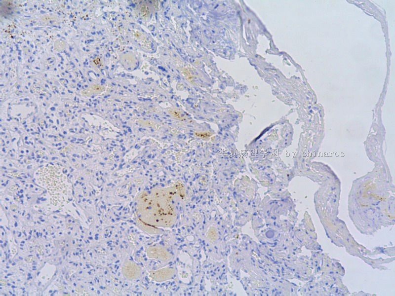
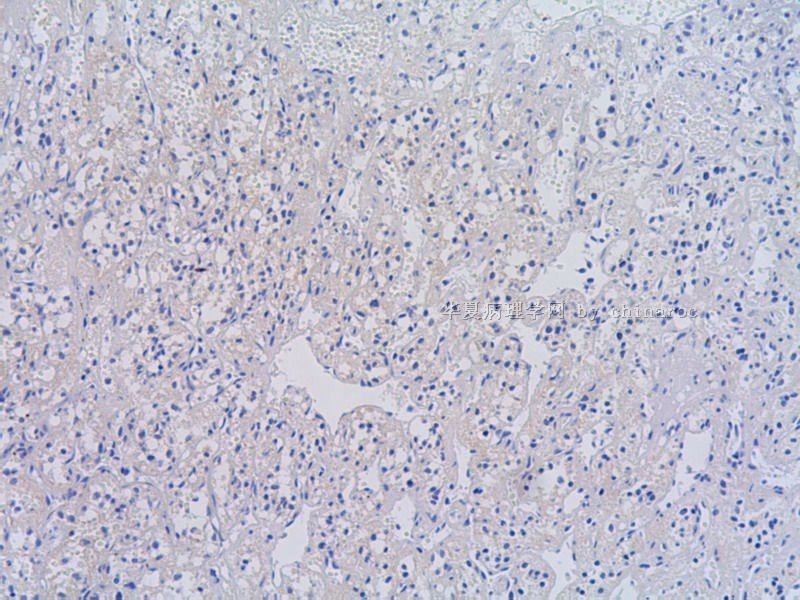
PR and EMA were negative, so meningioma can be safely ruled out. I am not sure how to interpret EGFR staining in this case. EMA for renal cell carcinoma is usually positive. It is certainly possible to have metastatic renal cell carcinoma in the brain, a usual case of which would display some degree of cytologic atypia and I did not see that in this case.
聞道有先後,術業有專攻


