| 图片: | |
|---|---|
| 名称: | |
| 描述: | |
- 35岁女性,卵巢肿物,是恶性吗?
| 以下是引用青青子矜在2009-12-24 19:13:00的发言:
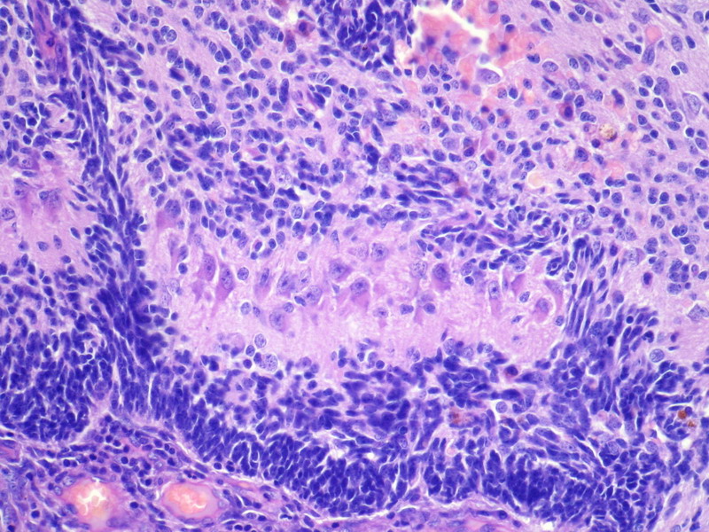
的确是很有意思的病例,多谢楼主分享! “35岁,卵巢肿物”虽然楼主未详细描述肿瘤巨检情况,但我想首先这些应该是肿瘤性成分而不是残存卵巢组织。 支持所示图片是不同发育阶段的脑组织,管状结构大部分是室管膜成分,是否有不成熟成分还要看全片情况。 如果没有其它成分,支持刘老师所说的“单胚层畸胎瘤”。 但新WHO分类中界定的“未成熟性畸胎瘤”专指原始神经管,分级也是根据其所占比例区分。至于畸胎瘤中其它恶性成分,则需要特殊说明,比如“畸胎瘤,伴有。。。成分,比例。。。”,但不叫“未成熟性畸胎瘤”,预后取决于伴随成分的恶性程度及所占比例。 不知道看法是否正确,希望各位专家老师们多指点。 期待下文 |


- If you have great talents, industry will improve them; if you have but moderate abilities, industry will supply their deficiency. 如果你很有天赋,勤勉会使其更加完美;如果你能力一般,勤勉会补足其缺陷。
-
本帖最后由 于 2009-12-25 01:25:00 编辑
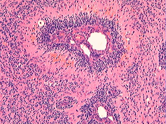
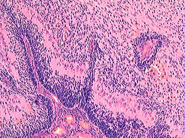
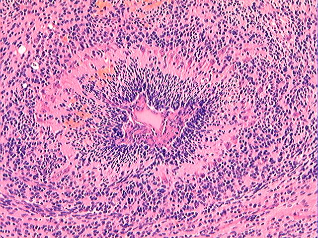
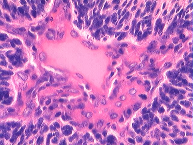
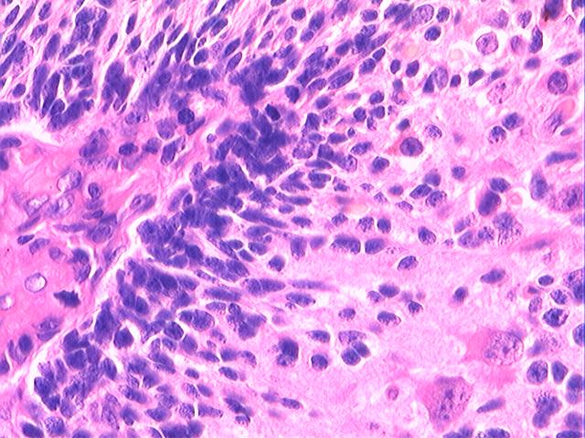
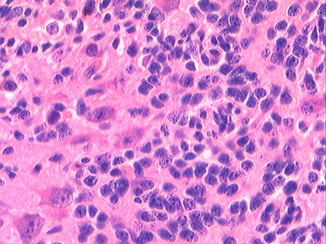
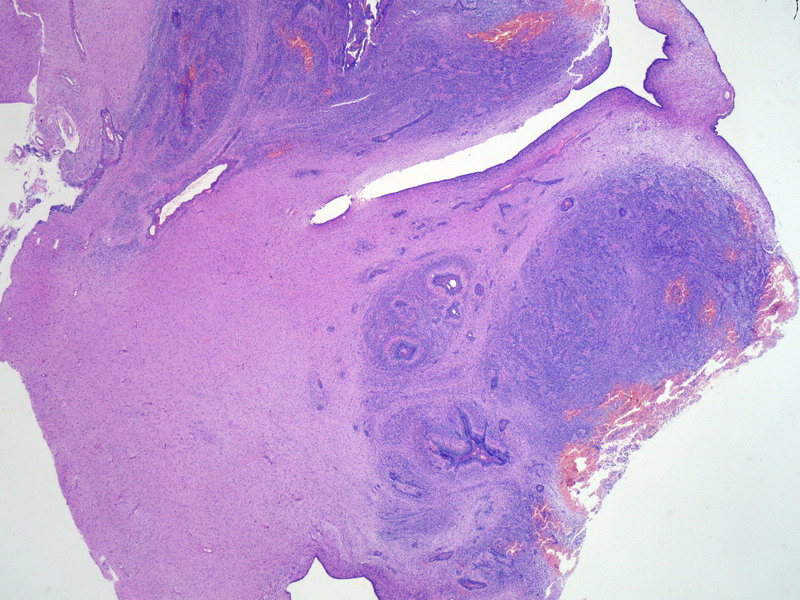
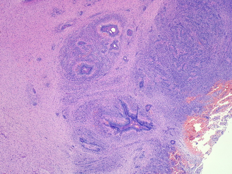
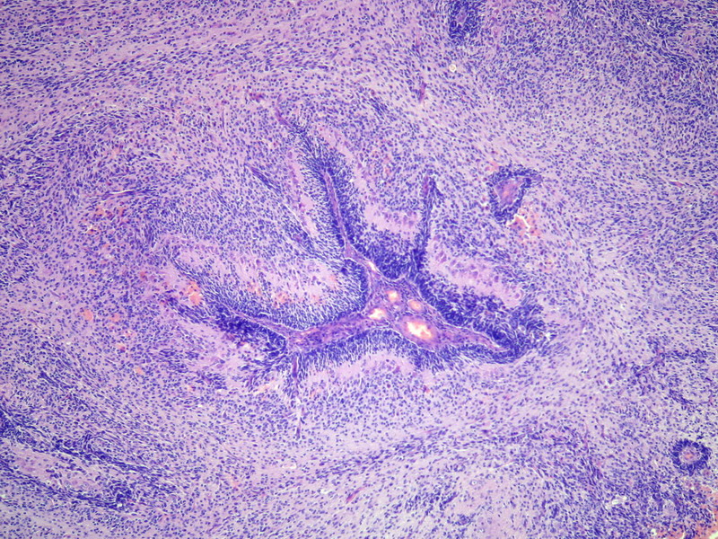
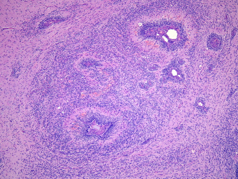
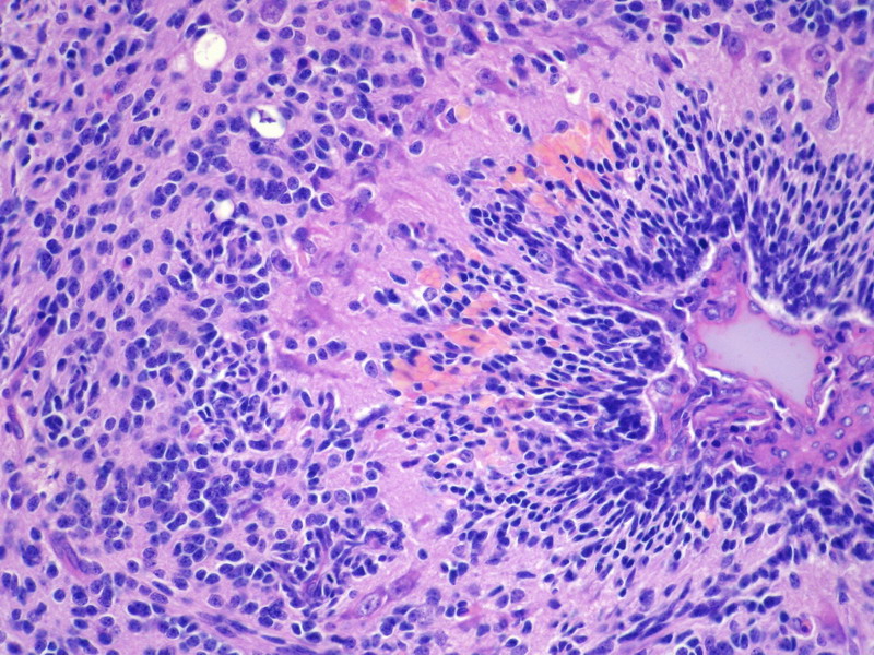
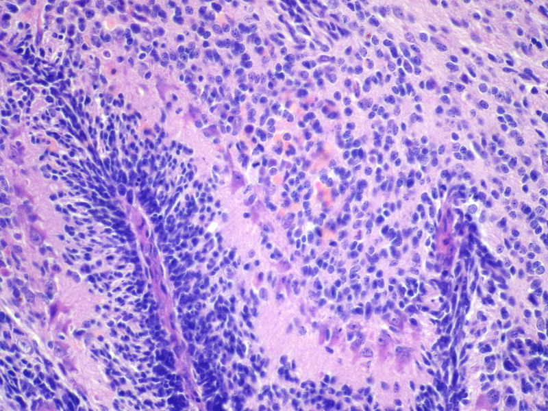
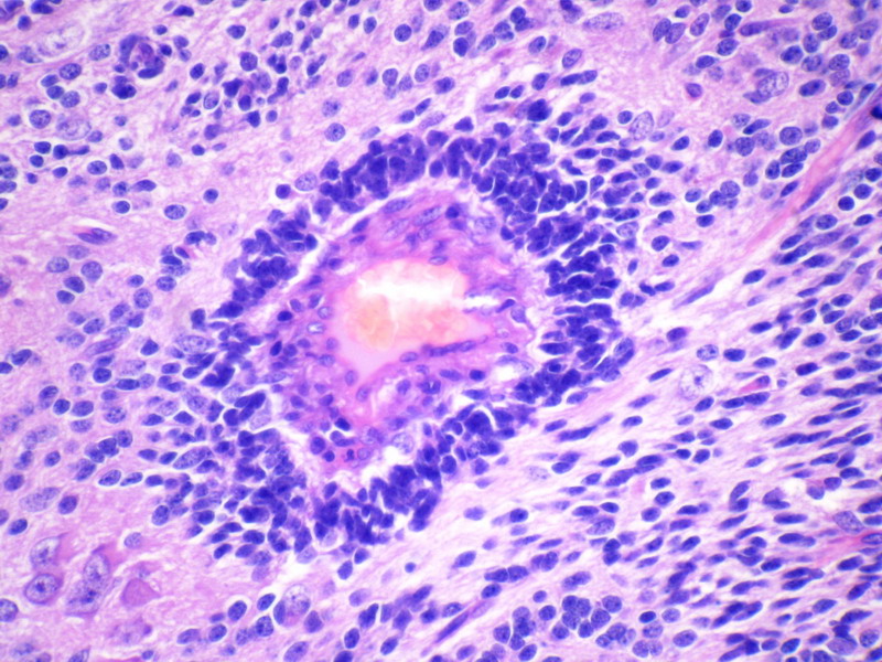
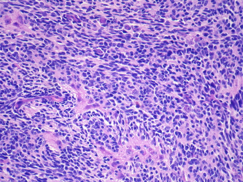
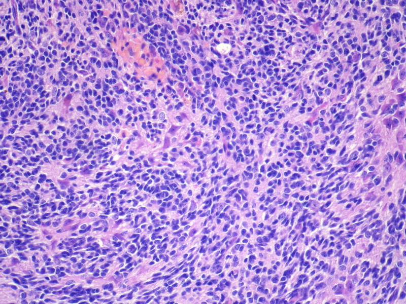
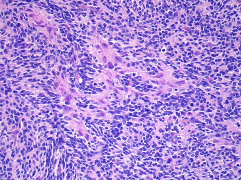
不知道这个肿块到底有多大,不知道这个肿块是否肉眼观有囊性结构。从提供的图片放大看,不是经典的神经管结构。但是,这管状结构的周围的确可见发育成熟的神经元和神经胶质。感兴趣的是,管状结构中央有较厚的管壁,有上皮内衬。似正在发育的中央导水管或室管膜样结构。问题是,围绕管状结构周围的深染的小短梭形或类圆形小细胞是什么细胞,还有一些区域可见成片的实性细胞聚集似乎将要形成“管状结构”。有可能就是未成熟的神经组织。
这可能是一个特例,虽然见不到经典的神经管。但从迹象看,可能是一些相对幼稚或曰胎儿型的神经组织,有类似于神经管分化的意义。因此,这可能是一个未成熟性畸胎瘤。多取材看看,如果没有皮肤及附件、原肠分化等结构存在,那可能是一个只有神经分化的单形性未成熟畸胎瘤。
未成熟畸胎瘤和恶性畸胎瘤绝不是一个相同的概念。未成熟是指胚胎发育的分化过程未达到成熟状态,如神经管,处在早期神经发育与分化阶段。将其分离在体外条件培养基中培养,是可以向成熟方向分化的。未成熟成分在一定意义上讲,是早期胎儿型的;而恶性畸胎瘤,其中含有至少有一种成分发生恶性转化,就如同经典的某组织起源的恶性肿瘤在畸胎瘤中。
以上议论,如有不妥,请鉴谅。

- 王军臣
| 以下是引用青青子矜在2009-12-24 19:13:00的发言:
的确是很有意思的病例,多谢楼主分享! “35岁,卵巢肿物”虽然楼主未详细描述肿瘤巨检情况,但我想首先这些应该是肿瘤性成分而不是残存卵巢组织。 支持所示图片是不同发育阶段的脑组织,管状结构大部分是室管膜成分,是否有不成熟成分还要看全片情况。 如果没有其它成分,支持刘老师所说的“单胚层畸胎瘤”。 但新WHO分类中界定的“未成熟性畸胎瘤”专指原始神经管,分级也是根据其所占比例区分。至于畸胎瘤中其它恶性成分,则需要特殊说明,比如“畸胎瘤,伴有。。。成分,比例。。。”,但不叫“未成熟性畸胎瘤”,预后取决于伴随成分的恶性程度及所占比例。 不知道看法是否正确,希望各位专家老师们多指点。 期待下文 |
青青子矜 did wonderful translation job! This is the exact environment I like to see more and more here. Everybody tosses their opinions based on morphologic analysis. There is no authority, but just different opinion and interpretations, as long as you can provide some in depth analysis. The point is not who is right at the end, but the differential diagnoses and how do you convince yourself and others to reach a logic and correct diagnosis.
Happy New Year to you all!

- 不坠青云之志,长怀赤子之心
的确是很有意思的病例,多谢楼主分享!
“35岁,卵巢肿物”虽然楼主未详细描述肿瘤巨检情况,但我想首先这些应该是肿瘤性成分而不是残存卵巢组织。
支持所示图片是不同发育阶段的脑组织,管状结构大部分是室管膜成分,是否有不成熟成分还要看全片情况。
如果没有其它成分,支持刘老师所说的“单胚层畸胎瘤”。
但新WHO分类中界定的“未成熟性畸胎瘤”专指原始神经管,分级也是根据其所占比例区分。至于畸胎瘤中其它恶性成分,则需要特殊说明,比如“畸胎瘤,伴有。。。成分,比例。。。”,但不叫“未成熟性畸胎瘤”,预后取决于伴随成分的恶性程度及所占比例。
不知道看法是否正确,希望各位专家老师们多指点。
期待下文
| 以下是引用杨斌在2009-12-24 13:03:00的发言:
Chengquan, no offend. You may be right. But it is so unusual to see an isolated few 'immature neuronal element" in such a clean back ground with no other "teratomatous" changes or components. It is so challenging to render right diagnosis without seeing the whole slide and whole pictures. The view of this case is so focused with lot of high magnifications. I feel there is no way I can call this an "immature teratoma" only based on these pictures. Just my two pennies to arouse further discussion here. Merry Christmas! |
澄泉,无意冒犯!你可能是对的,但如此干净背景下仅见孤立的少量“不成熟神经成分”而无其它“畸胎瘤”改变或成分的确是太罕见了。不看全部切片和图片而要作出正确诊断太难了,这个病例的视野都集中在多数高倍镜下,我认为没办法仅凭这些图片诊断为“未成熟性畸胎瘤”。
这里抛砖引玉,意在唤起更多讨论。
圣诞快乐!
| 以下是引用cqzhao在2009-12-24 5:15:00的发言:
Interesting case. Bin, you have good differential diagnosis. However I cannot agree with you. I think these cells are neuronal component, but not sex cord stromal components. The key for this case to dianose immature teratoma or mature solid teratome depends on that the neuroal component is immature and mature. My feeling is that neuronal component is immature though I am not completely sure that. Need to check books to learn neuronal pathology or get consult from neuropathologists for final diagnosis. |
很有趣的病例!
斌,你作了很好的鉴别诊断,但我不同意你的观点。
我认为这些细胞是神经成分而不是性索间质,但关键是这个病例到底是诊断为未成熟性畸胎瘤还是实性成熟性畸胎瘤?这取决于那些神经成分是未成熟性还是成熟性。
我感觉那些神经成分是不成熟的,虽然我没有十分把握。
需要查阅一些神经病理学书或请神经病理学家会诊来做最终诊断。
| 以下是引用杨斌在2009-12-23 1:26:00的发言:
I am not convinced this is a neoplastic lesion. To me it appears more physiologic than pathologic. Most of photos show an atretic follicle with central vascular structure, a layer of atrophic and splindle granulosa cell layer and a somewhat lutinized theca interna layer which is merging with ovarian stroma. The clue is the prominent eosinophilic and hyalinized so-called "glassy membrane" band in between dark spindle granulosa layer and lutinized theca interna. There are scattered lutinized thecal cells in theca interna with ample pink cytoplasm which are usually seen in childhood or pregnancy. I wonder if this patient has recent pregnancy or abortion history. Please let us know that. In any sense, I will be reluctant to make neoplastic diagnosis based on these photos alone. On the other hand, there is a large homogenous eosinophilic area in low power photos, but not showing high power pictures. I am curious of the changes in that area and want to make sure we do not miss a spindle cell neoplasm such as ovarian fibroma. Please provide high power photos of those. My guess is that It is most likely just nothing but an area with spindle ovarian stroma. |
不确定这是个肿瘤性病变,我认为生理性表现胜过病理性病变。多数照片显示为具中央脉管结构的闭锁卵泡,一层营养层加上梭形粒层细胞以及被卵巢间质吸收融合的某些黄素化卵泡膜内层。诊断线索是明显的嗜酸性胞质和位于暗梭形颗粒细胞层和黄素化卵泡膜之间的透明化的所谓 “透明膜”带,在卵泡膜内层有散在黄素化卵泡膜细胞,胞质淡粉染,这种细胞常见于儿童期或孕期。病人是否有近期怀孕或流产史?请让我们知道。感觉上,我很不愿意仅凭这些照片就诊断为肿瘤。
另一方面,低倍镜下见很多同质嗜酸性细胞区域,但并未显示高倍镜特征,我对这些区域变化很感兴趣,需要确定是否漏诊了象卵巢纤维瘤之类的梭形细胞肿瘤。
请提供这些区域的高倍镜图片,我猜测很可能没问题,只是梭形细胞卵巢间质。
Chengquan, no offend. You may be right. But it is so unusual to see an isolated few 'immature neuronal element" in such a clean back ground with no other "teratomatous" changes or components. It is so challenging to render right diagnosis without seeing the whole slide and whole pictures. The view of this case is so focused with lot of high magnifications. I feel there is no way I can call this an "immature teratoma" only based on these pictures. Just my two pennies to arouse further discussion here.
Merry Christmas!

- 不坠青云之志,长怀赤子之心
Interesting case.
Bin, you have good differential diagnosis. However I cannot agree with you. I think these cells are neuronal component, but not sex cord stromal components.
The key for this case to dianose immature teratoma or mature solid teratome depends on that the neuroal component is immature and mature.
My feeling is that neuronal component is immature though I am not completely sure that. Need to check books to learn neuronal pathology or get consult from neuropathologists for final diagnosis.
I am not convinced this is a neoplastic lesion. To me it appears more physiologic than pathologic. Most of photos show an atretic follicle with central vascular structure, a layer of atrophic and splindle granulosa cell layer and a somewhat lutinized theca interna layer which is merging with ovarian stroma. The clue is the prominent eosinophilic and hyalinized so-called "glassy membrane" band in between dark spindle granulosa layer and lutinized theca interna. There are scattered lutinized thecal cells in theca interna with ample pink cytoplasm which are usually seen in childhood or pregnancy. I wonder if this patient has recent pregnancy or abortion history. Please let us know that. In any sense, I will be reluctant to make neoplastic diagnosis based on these photos alone.
On the other hand, there is a large homogenous eosinophilic area in low power photos, but not showing high power pictures. I am curious of the changes in that area and want to make sure we do not miss a spindle cell neoplasm such as ovarian fibroma. Please provide high power photos of those. My guess is that It is most likely just nothing but an area with spindle ovarian stroma.

- 不坠青云之志,长怀赤子之心
| 以下是引用liu_aijun在2009-12-20 19:47:00的发言: 图片显示的全部是不同发育阶段的脑组织。考虑为单胚层畸胎瘤。 |
这些图片非常漂亮 
如果这些图片代表的是肿瘤全部或绝大部分。可以确诊为单胚层畸胎瘤。
今天专门请教了我们科搞神经病理的桂秋萍教授,那些围绕血管腔的结构是模拟大脑的胚胎发育过程,相当于大脑皮层,以及蛛网膜下腔。所以即不是神经管,也不是室管膜结构。
这一例诊断不成熟畸胎瘤,不是依据原始神经管成分。是单胚层畸胎瘤,但不是室管膜瘤,而是低倍视野中有一区域由成片的小细胞构成,相当于神经母细胞瘤。
好例子,谢谢分享。

- If you have great talents, industry will improve them; if you have but moderate abilities, industry will supply their deficiency. 如果你很有天赋,勤勉会使其更加完美;如果你能力一般,勤勉会补足其缺陷。

