| 图片: | |
|---|---|
| 名称: | |
| 描述: | |
- Inguinal lymphadenopathy
-
panzenggang 离线
- 帖子:189
- 粉蓝豆:480
- 经验:246
- 注册时间:2008-01-09
- 加关注 | 发消息
| 姓 名: | ××× | 性别: | Male | 年龄: | 70 years |
| 标本名称: | Inguinal lymph node | ||||
| 简要病史: | 70-year-old male, multiple inguinal lymphadenopathies with largest one up to 2.0 cm. No other clinial abnormalities. | ||||
| 肉眼检查: | Needle biopsy of inguinal lymph node. | ||||
名称:图1
描述:图1
名称:图2
描述:图2
名称:图3
描述:图3
名称:图4
描述:图4
-
panzenggang 离线
- 帖子:189
- 粉蓝豆:480
- 经验:246
- 注册时间:2008-01-09
- 加关注 | 发消息
-
本帖最后由 于 2009-12-23 10:44:00 编辑
-
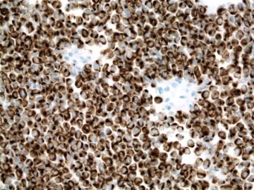
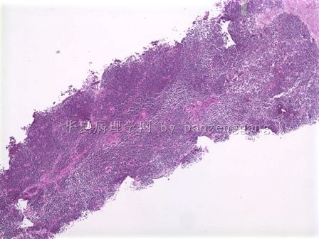
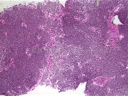
Diagnosis: metastatic Merkel cell carcinoma.
-
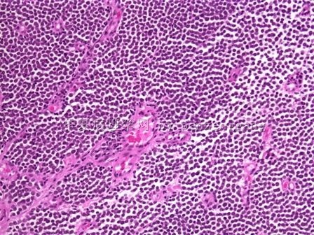
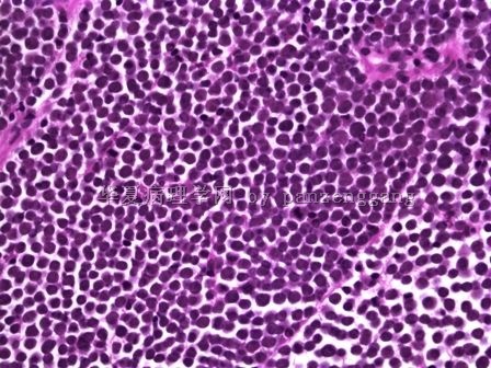
Microscopic description: The tumor consists of monotonous, small, round and blue cells with very high N/C ratio and evenly dispersed chromatin. The nucleolus is inconspicuous. The tumor cells are dyscohesive with focal vaguely nodular pattern. Abundant mitotic figures are present.
-
Differential diagnosis:
-
T-ALL: young age, mediastinal mass, TdT+, CD3+.
-
B-ALL: young age, more common as leukemia form, TdT+, CD10+, CD20+.
-
Metastatic small cell carcinoma for the lung: CK20-, CK5/6+, TTF+ (50%), CEA+/-.
-
Small cell melanoma: S100+, MITF+. MART1+, HMB45+, CK20-.
-
PNET: CD99+, Vimentin+, CK20-.
-
Merkel cell carcinoma: CK20+, CK5/6-, p63-, TTF1-, CEA-, S100-, vimentin-, CD45-, CD117+, Synaptophysin+, chromogranin A+.
-
-
Tumor
PanCK CK20 CK5/6 P63 TTF1 CD99 CEA S100 Melan-A
Vimentin CD45
Merkel cell carcinoma + + - - - +/- - - - - - Metastatic small cell carcinoma (lung) + - - - + +/- +/- - - - - Small cell squamous carcinoma + - + + - - - - - +/- - Small cell eccrine carcinoma + - - - - - +/- +/- - +/- - Small cell melanoma - - - - - +/- - + + + - Primitive neuroectodermal tumor +/- - - - - + - - - + - Non-Hodgkin's Lymphoma - - - - - +/- - - - +/- +
|
Summarized by Zenggang Pan, MD, PhD For more detail, please visit: http://www.enjoypath.com/hp/hp-073/hp-073.htm |
-
panzenggang 离线
- 帖子:189
- 粉蓝豆:480
- 经验:246
- 注册时间:2008-01-09
- 加关注 | 发消息
-
本帖最后由 于 2009-12-13 15:37:00 编辑
(Sorry I cannot type Chinese, and I can’t install Chinese since my Department gave me the computer during residency training)
The HE stain was OK.
Actually when I first saw this case, I had the same question: why the staining is so dark and weird? The nuclei are so dark and uniform, almost like ground glass pattern. could that be due to fixation or other artifact? Probably not, since it was a small needle biopsy and should be very easy to fix, and I did not see any signs of artifact. Anyway, the nuclei are really dark with very homogeneously fine chromatin.
Dr. XLJin8 gave very good differential diagnoses. However, we can probably rule out several of them just based on HE:
1). Plasma cell myeloma: larger cells with more cytoplasm and more clumped chromatin;
2). T/NK: moderate clear cytoplasm, and more irregular nuclei, classic vascular proliferation with inflammatory cells in the background.
3). Myeloid tumor: medium to large cells with some cytoplasm depending upon the stage of differentiation, but the chromatin should be very open with prominent nucleoli if in the blast stage, or should show some evidence of differentiation if in other stage.
4). Small round blue cell tumor: most likely, but again there are several entities, including small cell carcinoma of the lung, PNET, ALL, and small cell melanoma. But what most puzzled me was the nuclei: dark and homogeneous, more fine than "salt and pepper".
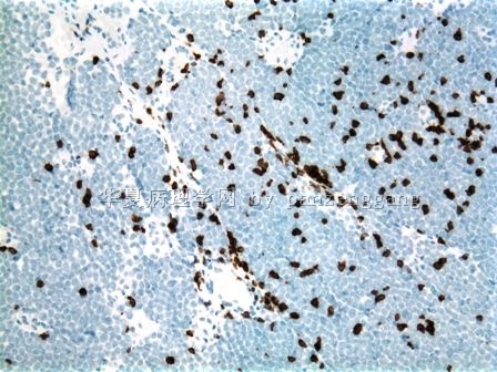
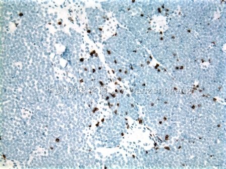
This case was showed in our Department QA meeting. My attending showed us the CD3 and CD79a first. When I saw both stainings were negative, I suddenly thought of a tumor with this kind of dark and fine chromatin, except the fact that it may have organoid growth pattern which didn't show in this lymph node. I asked her: do you have CK20? She was very surprised. Actually, at the time, I just started collecting the cases of "metastatic tumors in the lymph node mimicing lymphomas", and it was not surprising that I had this idea.
IPX: CK 20+ , as shown below. He had no skin lesion.
