| 图片: | |
|---|---|
| 名称: | |
| 描述: | |
- Esophageal mass
-
panzenggang 离线
- 帖子:189
- 粉蓝豆:480
- 经验:246
- 注册时间:2008-01-09
- 加关注 | 发消息
| 姓 名: | ××× | 性别: | Male | 年龄: | 66 years |
| 标本名称: | Esophageal mass | ||||
| 简要病史: | 66 year-old male, progressive dysphagia for 2-3 years. Image studies revealed an esophageal mass located at 34-37cm from the incisors | ||||
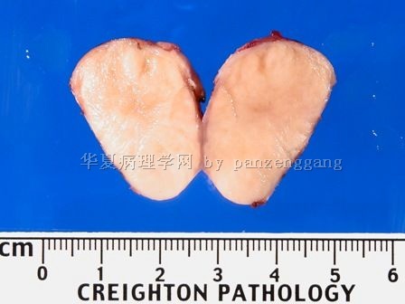
| 肉眼检查: | The specimen consists of one round, tan-gray nodule measuring 5.0 x 2.5 x 2.0 cm. The outer surface is smooth. Sections through the specimen reveals a homogeneously pale-yellow cut surface without any hemorrhage or necrosis. | ||||
名称:图1
描述:图1
名称:图2
描述:图2
名称:图3
描述:图3
名称:图4
描述:图4
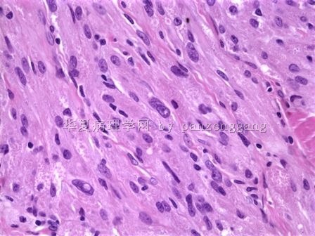
名称:图5
描述:图5
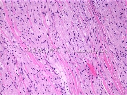
名称:图6
描述:图6
标签:
×参考诊断
Final Diagnosis:
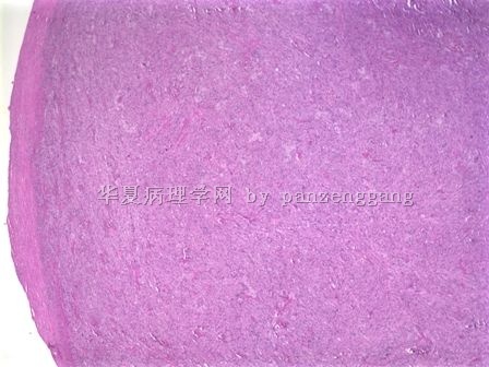
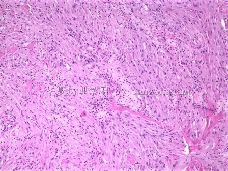
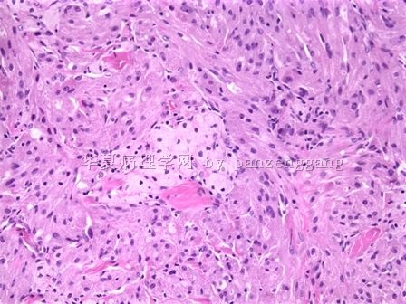
Granular cell tumor


