| 图片: | |
|---|---|
| 名称: | |
| 描述: | |
- Omental masses(网膜肿块)
| 姓 名: | ××× | 性别: | female(女) | 年龄: | 66 |
| 标本名称: | Omental biopsy(网膜活检) | ||||
| 简要病史: | |||||
| 肉眼检查: | |||||
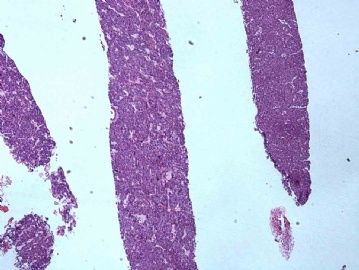
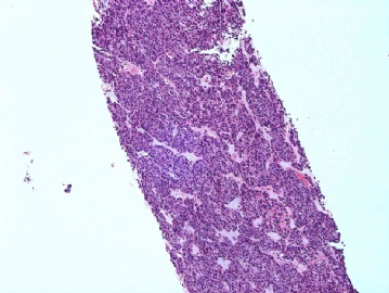
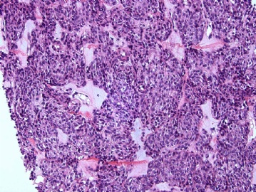
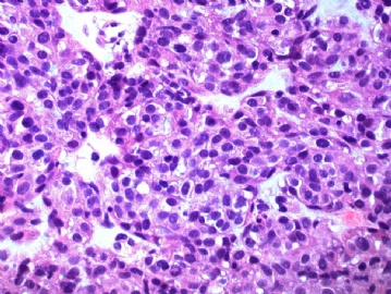
This is a 66-year-old lady with multiple omental masses. The gynecologist wants to know if this is a tumor of the GYN tract? What's your differential diagnosis?
(66岁女性,多发性网膜肿块。妇科要求知道这是否是女性生殖道肿瘤?你的鉴别诊断?__abin译)
-
本帖最后由 于 2011-03-11 10:03:00 编辑
-
本帖最后由 于 2009-10-21 21:17:00 编辑
谢谢大家的讨论。Abin, you are the first one to raise the possibility of GIST from the extra-gastrointestinal tract, job well-done! 96298: you posted the exact abstract that I would like to post also, which is an excellent paper on omental GIST. Thank you!
This is a case of extra-gastrointestinal GISTs presented as omental masses (NOT endometrial mass). Yes, I did CD34 and it is focally positive, not as strong as CD117. Now, this case was sent to an outside institution for mutational analysis as suggested by the above paper. This is the second case I had this year of an omental GIST, just like to share with you
(大致翻译:
abin第一个提出胃肠道外GIST的可能性。96298提供了准确的文献摘要,这也是我想做的,这是一篇关于网膜GIST的好文献。谢谢你们!
这是一例胃肠道外GIST,表现为网膜肿块(不是内膜肿块)。CD34局灶阳性,没有CD117表达那么强。现在本例送往院外研究机构做突变分析,如文献所建议。这是今年我遇到的第二例网膜GIST,很高兴与大家分享。
abin译)
-
本帖最后由 于 2009-10-20 18:14:00 编辑
The American Journal of Surgical Pathology: September2009 Volume33 Issue9 pp1267-1275
Gastrointestinal Stromal Tumors Presenting as Omental Masses-A Clinicopathologic Analysis of 95 Cases
Abstract
Gastrointestinal stromal tumors (GISTs), generally KIT-positive and KIT/PDGFRA mutation-driven mesenchymal neoplasms, most commonly originate from the stomach or small intestine, but in rare examples they involve the omentum. In this study, we analyzed 95 GISTs surgically designated as the omental masses. These tumors occurred in 49 males and 46 females with a median age of 60 years (range: 27 to 88 y). They formed single (n=51) or multiple masses (n=39); 5 cases were equivocal in this respect. Of the single tumors, 21 had no evidence of gastrointestinal tract involvement, 25 were attached to stomach, and 3 were attached to small intestine. Clinicopathologic parameters and prognosis of the 2 former groups were similar. Single tumor cases showed a median mitotic count of 2/50 HPFs and median tumor size was 14 cm. Their histologic features were similar to gastric GISTs in 22 cases, and to small intestinal GISTs in 6 cases. These tumors were KIT positive 38/41, CD34 positive 20/33, 8 had PDGFRA mutations, and 6 had KIT exon 11 mutations. The median survival was 129 months (range: 0 to 397 mo) and 14 patients were alive at the end of follow-up. Multiple tumor cases showed median mitotic count of 14/50 HPFs and the main tumor median size was 16 cm. The histologic features were similar to small intestinal GISTs in 21 cases and to gastric GISTs in 7 cases; small intestinal attachment or history of a previous small intestinal GIST were noted in 5 cases, whereas no tumor was attached to stomach. The multiple GISTs were KIT positive 23/24, CD34 positive 7/21, and 5 had KIT exon 11 mutations, 3 had KIT exon 9 mutations, and 2 had PDGFRA mutations. The median survival was for 8 months and all patients died. Omental GISTs are clinicopathologically heterogenous. Patients with solitary tumors usually have gastric GIST-like morphology and a better prognosis than those with multiple tumors, whose tumor usually has small intestinal GIST-like histology. Omental GISTs unattached to gastrointestinal tract often resemble gastric GISTs suggesting that they may be gastric GISTs directly extending or parasitically attached into the omentum, whereas multiple omental GISTs more often resemble small intestinal GISTs suggesting that they may be metastatic or detached from this source. KIT positive Cajal cells were not found in normal omental tissues failing to support the presence of these ancestral cells for GIST in the omentum.
| 以下是引用96298在2009-10-19 23:41:00的发言:
看来是GIST转移到子宫内膜了,真有点不可思议。 Nice case! |
不是内膜活检,是网膜活检。是否楼主笔误?
根据上述形态及免疫组化,可以确诊GIST,如果胃肠确实没原发灶,就是胃肠外GIST了。这种情况并非罕见。
谢谢陈博士提供穿刺病例,随着微创技术的广泛开展,这样的例子越来越多,病理医生的压力也越来越大了。

- If you have great talents, industry will improve them; if you have but moderate abilities, industry will supply their deficiency. 如果你很有天赋,勤勉会使其更加完美;如果你能力一般,勤勉会补足其缺陷。
-
本帖最后由 于 2009-10-21 21:09:00 编辑
I also did the following immunos: CD10 negative, HMB45 negative, Inhibin negative, synaptophysin and chromogranin negative. The only stain that is strongly positive is CD117 (c-kit). Any thoughts?
(我也做了以下免疫组化:CD10-,HMB45-,Inhibin-,Syn-,CgA-。仅CD117 (c-kit)强阳性。有何考虑?__abin译)
-
本帖最后由 于 2009-10-21 21:07:00 编辑
Thank you abin for the translation and discussion. I did all the cytokins and mesothelial markers (AE1/AE3, CK7, Cam5.2, EMA, CK5/6, Calretinin, and WT-1), they are all negative. What should I do?
(谢谢abin翻译并讨论。我做了CK和间皮标记物(AE1/AE3, CK7, Cam5.2, EMA, CK5/6, Calretinin, and WT-1),它们全部阴性。下一步我该怎么办?__abin译)
翻译了请大家讨论。我先抛个砖头:
上皮样细胞形成实性\乳头状结构,高倍可见一些小圆孔,这可能是内膜样癌的特征。多发性,更可能是转移来的。
首先考虑内膜样腺癌(来自子宫或卵巢)。
鉴别:浆液性癌(来自子宫或卵巢),性索-间质肿瘤(来自卵巢,可能性不大),上皮样间皮瘤(腹膜原发或胸膜等其它部位转移),GIST(来自胃肠道或腹膜原发,可能性不大)。
order IHC:ER,PR,CK,VIM,calretinin,CK5/6,CD117。
影像学资料有助于诊断。

华夏病理/粉蓝医疗
为基层医院病理科提供全面解决方案,
努力让人人享有便捷准确可靠的病理诊断服务。


