| 图片: | |
|---|---|
| 名称: | |
| 描述: | |
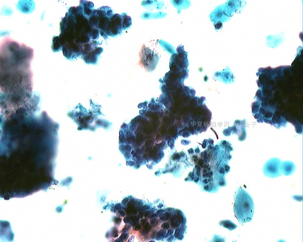
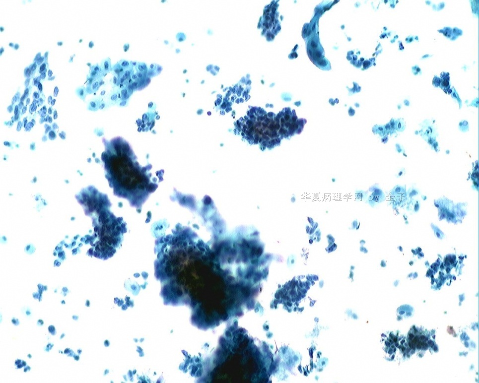
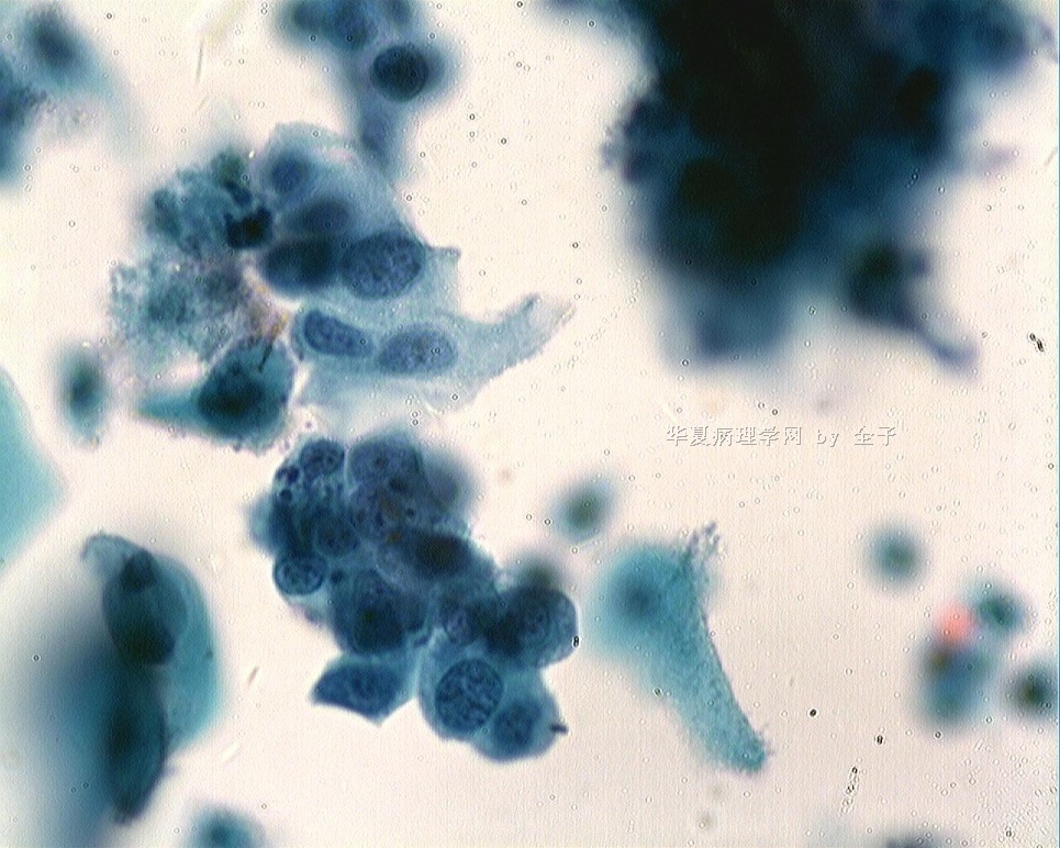
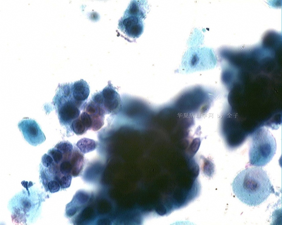
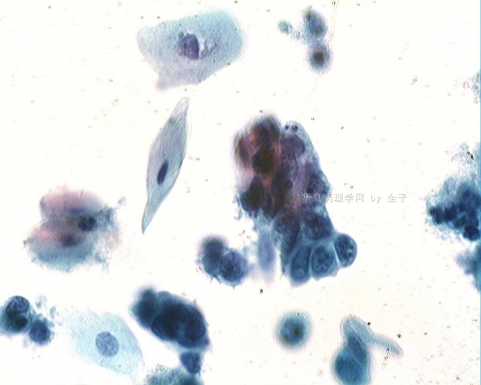
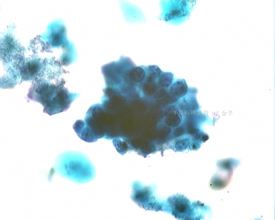
- 51y宫颈液基
| 以下是引用掌心0164在2009-9-9 19:15:00的发言:
这可是一个复杂的而少见的病例;走弯路纯属于正常。在宫颈出现腺鳞的病变难以鉴别的时候。如果年龄年轻;绝经前;细胞学大多考虑鳞状上皮的病变为主。但是绝经之后;就刚好相反;考虑腺病变为主。这类正好是一个刚绝经的病类;正好有同时具备了腺鳞病变;所以走弯路纯属于正常。我们只有在这种病案中不停总结中进步。有好病例请全子姐姐多发上来给我们好好学习学习!谢谢! |
刚看到一篇文献,楼主的这个病例也许可以归为“腺样基底细胞癌”。全子:你对照看看是否符合呢?我来不及翻译。或者你个人想法???
Adenoid basal lesions of the uterine cervix: evolving terminology
and clinicopathological concepts
Michael J Russell1,2 and Oluwole Fadare*1,3
The epithelial proliferations that are designated adenoid basal carcinoma (ABC) in the current
classification from the World Health Organization represent <1% of all cervical malignancies. These
lesions may be associated, and occasionally show morphologic transitions with, conventional
cervical malignancies. The determination of the precise frequency with which these so-called ABCs
show this association is hampered by the inherent selection bias in the reported cases. However,
this frequency appears to be substantial (>15%). The biologic course of ABCs that are associated
with separate malignancies is largely dependent on the clinicopathologic parameters of the
associated malignancies. Morphologically pure lesions, in contrast, have largely been associated with
favorable patient outcomes, as none of the 66 reported patients have experienced tumor
recurrence, metastases or tumor-associated death, irrespective of the modality of treatment.
Although the finding of genome integrated high-risk human papillomavirus (HPV) types and p53
alterations in adenoid basal lesions (ABL) argue in support of their neoplastic nature, we identified
no lines evidence that suggest an inherent malignancy for morphologically pure lesions. The finding
of morphologic transitions between ABLs and conventional malignancies and shared HPV types in
these areas, suggest that ABLs have some malignant potential. However, the precise magnitude of
this potential is not readily quantifiable and should not dictate the management of morphologically
pure lesions that are entirely evaluable. ABLs continue to occupy a unique position in human
oncology in which the term carcinoma (without an in-situ suffix) is applied to a tumor that has not
been shown to recur, metastasize or cause death. We concur with a previous proposal that the
term ABC should be discarded and replaced with Adenoid Basal Epithelioma (ABE). In our opinion,
there is insufficient evidence at present time to expose patients with morphologically pure lesions
to the ominous implications – social, psychological, medical, financial – of a "carcinoma" diagnosis.
Morphologically impure lesions should not be designated ABC or ABE. Furthermore, given the
uncertainties regarding the frequency with which ABE are associated with separate malignancies,
we suggest that the ABE designation only be applied when the tumor in question is entirely
evaluable e.g in a hysterectomy specimen or in an excisional biopsy with negative margins.
Otherwise, the generic designation Adenoid Basal Tumor is preferable. This approach strikes an
appropriate balance between the need to prevent over-treatment of pure lesions on one hand, and the need to ensure that the lesions are indeed pure on the other.
| 以下是引用xymady在2009-9-11 8:42:00的发言: 青青子矜,您太敬业啦,真正的学者风范和求真精神,细胞学园地有您们这些奉献者和支持者能不火乎?呵呵。另外提一点建议给您和掌心0164,请不要称呼我为“师傅”行吗,我那一点点皮毛功夫在网络上各位老师面前实在是班门弄斧,我实在是觉得愧不敢当/无地自容,请谅解,说得不妥之处望海涵,呵呵! |
马老师:您不要太谦虚,我们都认为您是看细胞的实干家,那是没的说,您的学生也是遍布大江南北。我那两手都是从您那里学来的呢。当然还要不断学习才行,否则出去还真不敢自称是您的学生,呵呵!
您要不认我和掌心啊,我就 
刚刚才得意没几天呢。。。
以后您就领着我们一道进步吧
| 以下是引用掌心0164在2009-9-9 16:20:00的发言:
老师是高手,请您多来我们这里指点! 高手拉低手,大家一起走! |

