| 图片: | |
|---|---|
| 名称: | |
| 描述: | |
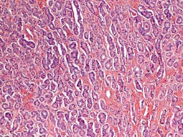
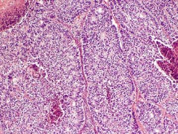
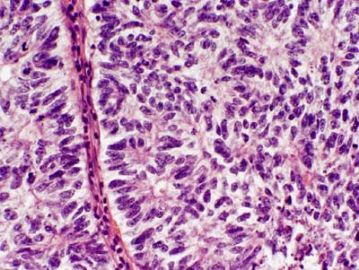
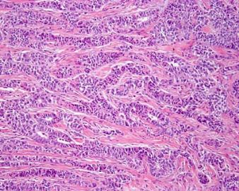
- 卵巢Sertoliform (or sex cord-like) variant of 子宫内膜样癌 (cqz-3)
说说我的浅显的看法:
60岁,单侧,10cm;细胞梭形,界限不清,染色质致密,细胞条索状位于间质背景中;图1、2细节不明显,但是形态符合Call-Exner小体。因此首先考虑颗粒细胞瘤。
但是真的诊断时候要注意寻找有无其他成分。再者如图所示成分占多少。即使有其他成分,甚至其他成分占多数,只要如图所示部分超过10%仍然诊断颗粒细胞瘤;如果无其他成分且如图所示部分很少,则诊断“伴有少量性索间质的纤维瘤”
还要值得注意的是图3中,核深染,大小不一,虽然部分细胞似有核沟,但不明显,且有核分裂及非典型核分裂(左下、左中、水印中h所覆盖的细胞等),因此应该想到未分化小细胞癌。
请各位老师批评指正……

- 赚点散碎银子养家,乐呵呵的穿衣吃饭
-
zxr48271117 离线
- 帖子:159
- 粉蓝豆:458
- 经验:577
- 注册时间:2009-07-30
- 加关注 | 发消息
-
zxr48271117 离线
- 帖子:159
- 粉蓝豆:458
- 经验:577
- 注册时间:2009-07-30
- 加关注 | 发消息
Agree. In addition, the upper left photo is typical adenocarcinoma, to my eyes
The upper right and lower left appear similar, same type with low and high power? The right lower looks different again. Could this be a collision tumor of different kinds?
Thank above a lot of very good analysis.
Summary above discussion:
Differential diagnosis (DDX):
1. Sex cord stromal tumor. If it is sex cord tumor, what type? granulosa tumor vs sertoli cell tumor.
2. Carcinoid tumor.
3. Primary epithelial tumor. What type? Serous, endometrioid
4. Metastatic tumor. From where?
I think this is good DDX. We must work with IHC stains. I have full panel of IHC results and will paste here next week.
