| 图片: | |
|---|---|
| 名称: | |
| 描述: | |
- 外阴Paget病和EMPD简要总结介绍(cqz-2)
Is Toker cells Precursor Cells???
n Toker cells are a feature of the interface between the epidermis and the epithelium of lactiferous ducts of the breast, ectopic breast tissue and MLG.
n Morphologically appear to be benign counterpart of malignant cells in PD.
n Supported by IHC staining
-
本帖最后由 于 2009-08-22 08:03:00 编辑
Mammary-like glands (MLG)
n Glands that normally occur in the interlabial sulcus between the labia minora and labia majora
n Histologic features of female breast and sweat glands
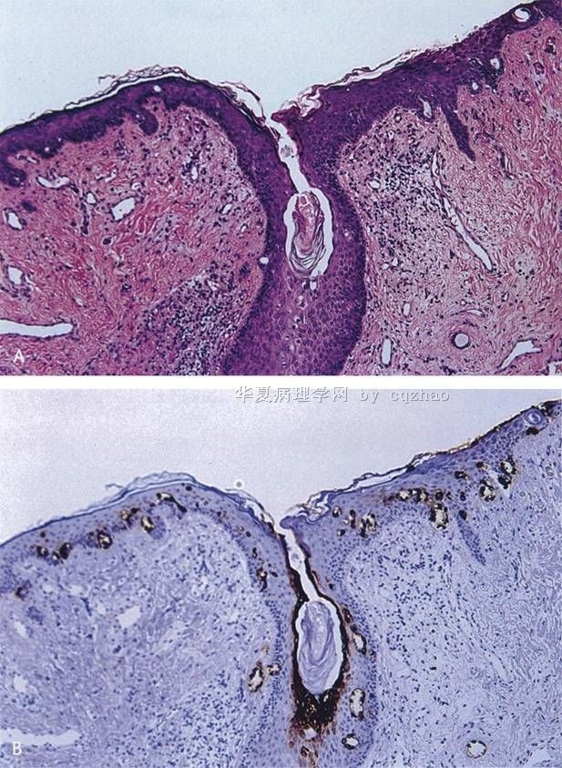
n A, Opening of MLG with formation of duct-like structures by hyperplastic Toker cells (H&E).
B, CK7 positivity in hyperplastic Toker cells (CK7).From: Willman: Am J Dermatopathol, Volume 27(3).June 2005.185-188

名称:图1
描述:图1
Primary EMPD
n Originate in the skin or apocrine sweat glands
n Not associated with distant adenocarcinoma
n Limited to the epidermis but may progress to invasive tumor, spreading to dermis, blood and lymphatics and potentially lethal metastases.
? Undifferentiated pluripotent cell of the epidermis or adnexa
? Toker cells of the vulvar epidermis
What are Toker cells?
n Intraepithelial cells with pale staining cytoplasm and bland cytologic features
n Located in the epidermis immediately adjacent to lactiferous ducts
n Considered a primitive germ cell of the lactiferous duct or inter-epithelial extension of lactiferous duct cells
n 10% normal nipples on H&E
83% normal nipples and 65% of accessory nipples with CK7 IHC
-
本帖最后由 于 2009-08-22 18:54:00 编辑
Glad to see above from Dr. Zhang and 青青子矜. I am a gynecologic/breast pathologist, but never do any research or write review about Paget's. Both of you did some research or wrote review articles in this area, so you know more detailed information. Thank for your expert's in-put.
I will continue to complete our resident's summary.
-
学习了,去年写了有关这方面的综述,把其中的结语摘录如下:
7.结语
在PD的各种变异型中,至少MPD的组织发生是有争议的。基于PC和其下的导管癌具有相同的黏液免疫表型,目前的研究支持亲表皮播散理论是其下有导管癌的MPD最重要的来源这种观点,而Toker细胞可能是部分其下没有乳腺癌的罕见MPD的来源。肛周的PD是特殊的,是仅表达MUC2的一组PD,提示MUC2做为识别可能来源于隐匿直肠癌的肛周PD的有用标记物。外阴PD的组织发生比MPD更复杂,乳腺样腺是外阴PD组织发生的另一侯选已被多位学者所推崇,应用粘蛋白基因做为一个新的标记重复验证了这种可能。外阴表皮内Bartholin腺与正常结构具有相同的粘蛋白表型,表皮内来自Bartholin腺或者MUC
PD是由几种奇特的病变实体构成的一组异质性病变,决定PD是表皮内还是表皮外来源对于治疗是最重要的,病理医生和临床医生都应该尽所有的努力检测所有的PD患者其下方是否有恶性肿瘤,黏蛋白基因表达在研究各种类型癌症的发生上是有帮助的,目前的研究显示MUC1几乎在所有的PD普遍表达,也可以用于筛选和确定表皮内PC的存在, MUC1比作为PC通用的上皮标记CK7要好,MUC1和CK7两者都出现在单一导管结构中(如小汗腺管、大汗腺管和Bartholin腺),提示单一导管或者他们的衍生物可能代表了EMPD各种亚型的起源。与直肠癌有关的肛周PD存在MUC2,在预知PD结肠来源方面MUC2似乎比CK20更特异。MUC
赵老师发起帖子来够吓人
谢谢分享这样的好病例。看了下很有启发,不一一翻译。
学生不才,最近刚刚写了篇文章中涉及到外阴,这里秀一下,希望对网友们能有点启发
Paget病 外阴是除乳腺外Paget病最常见的发生部位,大多数外阴Paget病是原发性的,但有5%是其它部位恶性病变(如膀胱尿路上皮癌、宫颈腺癌或结直肠腺癌等)的局部表现,此时外阴Paget病代表着这些恶性肿瘤的上皮内转移或扩散性病灶,因此辨别外阴Paget病是原发还是继发对临床治疗非常重要。多数研究证实原发性外阴Paget病的Paget细胞表达CK7、CEA和CAM5.2,大多数表达 GCDFP-15、HER-2和AR,ER和PR表达阴性。而来自于结直肠和和膀胱恶性肿瘤的外阴继发性Paget病通常表达该部位腺癌的免疫标记,如CK20、CDX2和MUC2阳性提示结直肠来源,而CK20和uroplakin Ⅲ阳性则为膀胱腺癌来源。最近有研究发现p63在外阴原发性Paget病与继发于膀胱尿路上皮癌的Paget病的鉴别诊断中具有价值,前者表达阴性,而后者表达阳性[8]。恶性黑色素瘤和外阴上皮内肿瘤(VIN)有时可有Paget样组织学构型,此时易误诊为外阴Paget病,但恶性黑色素瘤表达HMB45和S-100蛋白,而不表达CK7、CEA和GCDFP-15,Paget样VIN(Paget样鲍温病)CK7表达阴性。值得注意的是,正常皮肤内可含有CK7和CK20阳性的Merkel细胞,因此在诊断外阴Paget病时一定要结合形态学。
这样看来,赵老师这例我诊断---外阴原发性Paget病(CK7+、CEA+、CK5/6+、GCDFP-15弱+、HER-2+、p63大细胞--、CK20--、S-100--)
-
本帖最后由 于 2009-08-22 07:51:00 编辑
Pathogenesis of EMPD
n Unlike MPD, EMPD does not have a strong association with underlying adenocarcinoma (92-100% vs ~25%)
n Frequency of association of EMPD with underlying neoplasm varies with the site of the disease
n Two schools of thought:
n Paget's disease must arise from underlying neoplasm
n Paget's disease arises primarily in the epidermis
n Current theory is that there is dual origin of EMPDàPrimary vs. secondary
n Most cases of EMPD are primary
Two distinct types of EMPD
Type I- endodermal phenotype; associated with distant carcinomas, secondary
Common IHC phenotype
Type II- ectodermal/cutaneous; primary
-
本帖最后由 于 2009-08-22 07:48:00 编辑
Pathologic Features of PD
n Invasion of the epidermis by Paget cells (PC)
n Epithelial cells with abundant, clear cytoplasm
n Large, centrally situated nuclei with atypia and pleomorphism
n Signet ring aspect due to intracytoplasmic sialomucin
+ PAS, mucicarmine,
90% vs only 40% MPG
Pathologic Features of Extramammary PD
Lower epidermal layers, occasionally forming gland like structures with a central lumen
Outer epithelial layers of hair follicles or sweat gland excretory ductsEdematous dermis with dilated blood vessels and polymorphous inflammatory cell infiltrate
-
本帖最后由 于 2009-08-22 07:43:00 编辑
Vulvar EMPD
n 65% cases of EMPD
n Most cases are primary
n 4-17% associated with underlying adnexal neoplasm
n 11-20% associated with distant carcinoma
n Endometrial, endocervical, vaginal, bladder, colon, rectum, ovary, liver, gallbladder, skin
Differential Diagnosis
n Melanoma
n Bowen's disease
n Seborrheic or contact dermatitis
n Superficial fungal infections
n Basal cell carcinoma
n Flexural psoriasis
n Lichen sclerosus
-
本帖最后由 于 2009-08-22 07:46:00 编辑
Extramammary Paget's Disease (EMPD)
Epidemiology
n More rare than mammary Paget抯 disease (MPD)
n accounting for 6.5% of all cases
n MPD account for 1-4% of breast cancer
n 65-70 years of age, 90% over 50
n Female predominance of the vulvar localization (2% or less of primary vulvar neoplasms)
n No definite proof of a genetic predisposition
Clinical Features
n Insidious onset
n Pruritus
n Well-demarcated plaques may be separated by clinically normal skin that is pathologically involved
n Rarely, hypopigmented/hyperpigmented macules
n "underpants pattern erythema"due to lymphatic invasion and regional node enlargement and rapidly fatal distant metastases
n Diagnosis is often delayed up to 2 years until histological examination is performed secondary to chronic dermatosis not responding to local treatments.
Topographic Forms
n Perianal-20% EMPD
n Affects men and women equally, 63 yo
n Associated with adenocarcinoma of anus and rectum (~75%)
n Male Genitalia-14%
n Associated with underlying carcinoma of the ureter, bladder, prostate, testicle (11%)
n Axillary-more frequent in men, unilateral
n Exclusion of breast neoplasm is mandatory
n Ectopic-skin areas devoid of apocrine glands
n Chest, arms, fingers, eyelids, cheeks, scalp
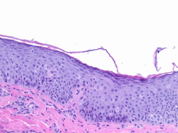
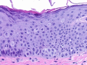
This is a vulvar Paget's/primary Paget's or called Extramammary Paget’s Disease
One of our first year residents made some summary about Paget's and showed in our morning study conference. I pasted the summary in the following.
As pathologists we should know lesions in more detailed. It is not enough to make dx only.


