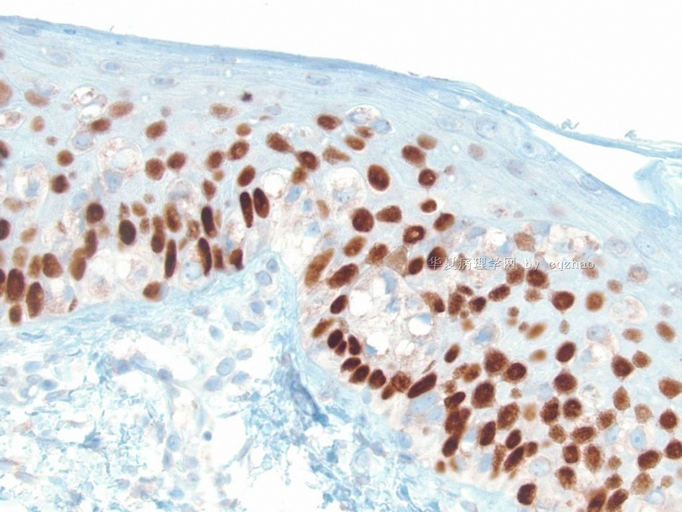
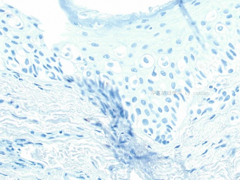
| 图片: | |
|---|---|
| 名称: | |
| 描述: | |
- 外阴Paget病和EMPD简要总结介绍(cqz-2)
| 以下是引用shandongzhang在2009-8-16 7:44:00的发言:
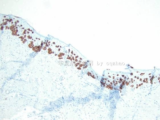
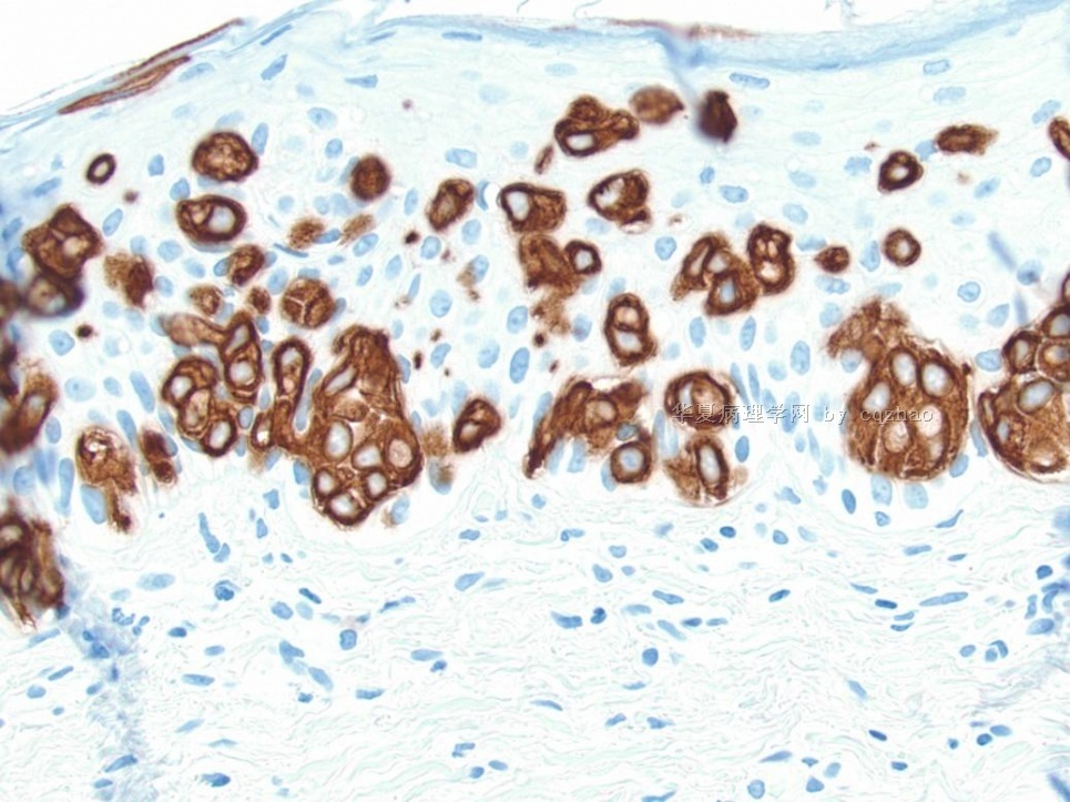
鉴别诊断: 1、表浅扩散型恶性黑色素瘤:异型细胞沿表皮、真皮交界处呈显著巢团状分布,无腺泡结构及细胞内黏液,且大部分病例黑色素标记物S-100蛋白、HMB-45、Melan-A和MART-1阳性,而CK8、MUC1、GCDFP-15等则多不表达。 2、Paget样Bowen病:常伴有鳞状上皮的增生、角化过度及角化不全、钉突延长,棘细胞不典型增生、黏液样变,角朊细胞核淡染、胞质透亮或呈空泡状,但无胞质内分泌现象,缺少大的异型细胞由基底层向表层播散的现象,免疫组化CAM5.2、GCDFP-15、Her-2标记阴性,虽然CK7阳性对PD的确诊有帮助,但要谨记,在Paget样Bowen病中也可能出现CK7阳性。 3、Paget样日光性角化病:具有特征性的组织学改变,由于角质形成细胞对摩擦形成的异常增生反应,通常在表皮上部出现异型细胞,但黏蛋白染色和CEA阴性可资区别。在大多数情况下,应用组织化学技术和免疫组化方法可资区别。 4、透明细胞丘疹病等 |
鉴别诊断非常详细,谢谢Dr.shandongzhang!
此例形态不典型,更倾向哪种呢?
-
Thank Dr. Zhang's good differential dx. When we see these kinds of lesions we must consider the differential dx. Three ddx including Paget's, Bowen, and melanoma should be in our mind immediately. The treatment and prognosis of these diseases can be huge different. We always do some stains for these lesions even though we can favor some dx based on H@E. You can be right in most of the time, but you will be wrong some time during your life long practice.
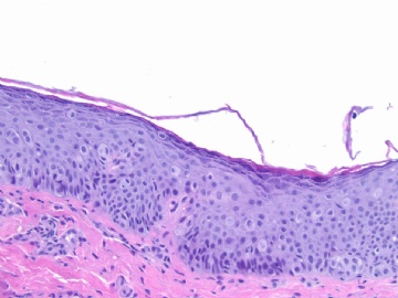
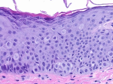
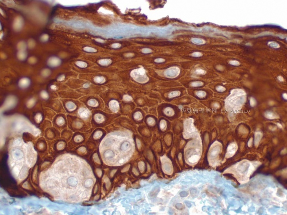
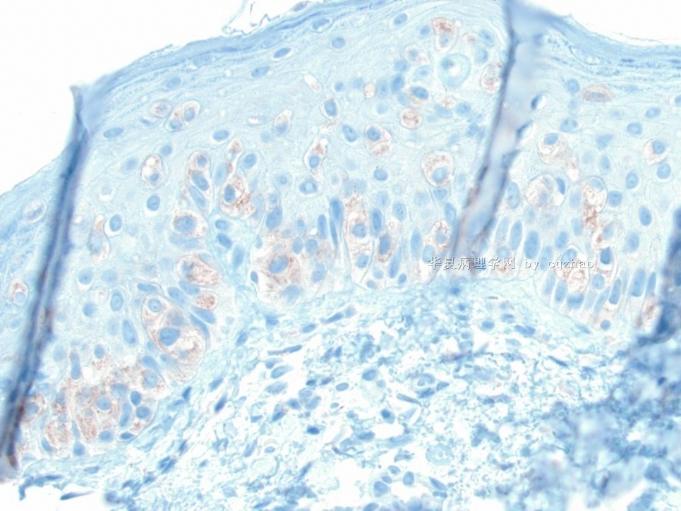
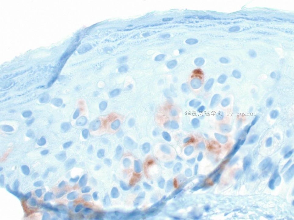
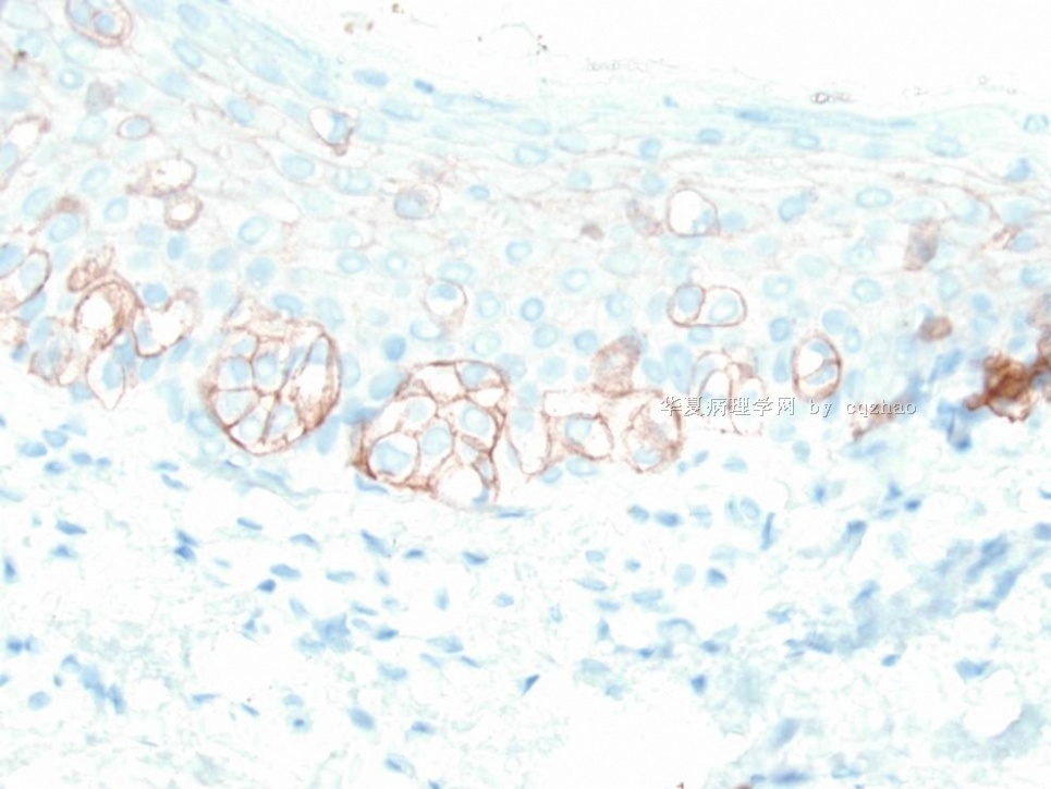
This is a vulvar Paget's/primary Paget's or called Extramammary Paget’s Disease
One of our first year residents made some summary about Paget's and showed in our morning study conference. I pasted the summary in the following.
As pathologists we should know lesions in more detailed. It is not enough to make dx only.
-
本帖最后由 于 2009-08-22 07:46:00 编辑
Extramammary Paget's Disease (EMPD)
Epidemiology
n More rare than mammary Paget抯 disease (MPD)
n accounting for 6.5% of all cases
n MPD account for 1-4% of breast cancer
n 65-70 years of age, 90% over 50
n Female predominance of the vulvar localization (2% or less of primary vulvar neoplasms)
n No definite proof of a genetic predisposition
Clinical Features
n Insidious onset
n Pruritus
n Well-demarcated plaques may be separated by clinically normal skin that is pathologically involved
n Rarely, hypopigmented/hyperpigmented macules
n "underpants pattern erythema"due to lymphatic invasion and regional node enlargement and rapidly fatal distant metastases
n Diagnosis is often delayed up to 2 years until histological examination is performed secondary to chronic dermatosis not responding to local treatments.
Topographic Forms
n Perianal-20% EMPD
n Affects men and women equally, 63 yo
n Associated with adenocarcinoma of anus and rectum (~75%)
n Male Genitalia-14%
n Associated with underlying carcinoma of the ureter, bladder, prostate, testicle (11%)
n Axillary-more frequent in men, unilateral
n Exclusion of breast neoplasm is mandatory
n Ectopic-skin areas devoid of apocrine glands
n Chest, arms, fingers, eyelids, cheeks, scalp
-
本帖最后由 于 2009-08-22 07:43:00 编辑
Vulvar EMPD
n 65% cases of EMPD
n Most cases are primary
n 4-17% associated with underlying adnexal neoplasm
n 11-20% associated with distant carcinoma
n Endometrial, endocervical, vaginal, bladder, colon, rectum, ovary, liver, gallbladder, skin
Differential Diagnosis
n Melanoma
n Bowen's disease
n Seborrheic or contact dermatitis
n Superficial fungal infections
n Basal cell carcinoma
n Flexural psoriasis
n Lichen sclerosus
-
本帖最后由 于 2009-08-22 07:48:00 编辑
Pathologic Features of PD
n Invasion of the epidermis by Paget cells (PC)
n Epithelial cells with abundant, clear cytoplasm
n Large, centrally situated nuclei with atypia and pleomorphism
n Signet ring aspect due to intracytoplasmic sialomucin
+ PAS, mucicarmine,
90% vs only 40% MPG
Pathologic Features of Extramammary PD
Lower epidermal layers, occasionally forming gland like structures with a central lumen
Outer epithelial layers of hair follicles or sweat gland excretory ductsEdematous dermis with dilated blood vessels and polymorphous inflammatory cell infiltrate
-
本帖最后由 于 2009-08-22 07:51:00 编辑
Pathogenesis of EMPD
n Unlike MPD, EMPD does not have a strong association with underlying adenocarcinoma (92-100% vs ~25%)
n Frequency of association of EMPD with underlying neoplasm varies with the site of the disease
n Two schools of thought:
n Paget's disease must arise from underlying neoplasm
n Paget's disease arises primarily in the epidermis
n Current theory is that there is dual origin of EMPDàPrimary vs. secondary
n Most cases of EMPD are primary
Two distinct types of EMPD
Type I- endodermal phenotype; associated with distant carcinomas, secondary
Common IHC phenotype
Type II- ectodermal/cutaneous; primary
