| 图片: | |
|---|---|
| 名称: | |
| 描述: | |
- 放疗后乳房(cutaneous)高级别血管肉瘤(cqz-23)
| 以下是引用zhanglei在2009-7-25 18:48:00的发言:
出乎意料之外的结果! 思维定式对我们影响太深了!结合病史,首先排除复发性癌的考虑是多数网友都会想到的,但镜下图像用哪种癌都无法解释,怎么办?我想以后我会想到放化疗后患者继发血管肉瘤的几率要大一些的,如果本例头脑中有一点这样的概念,再根据图像中肿瘤细胞与血管的密切程度或许能够考虑到血管肉瘤的诊断。 再次谢谢楼主提供的精彩病例! |

- 博学之,审问之,慎思之,明辨之,笃行之。
-
本帖最后由 于 2009-07-26 08:22:00 编辑
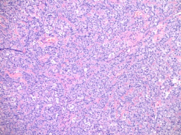
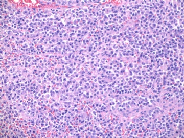
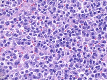
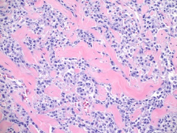
This biopsy speciment was diagnosed as high grade angiosarcoma and the pt had mastectomy. Paste here some photos from mastecetomy. I want to show you the high grade angiosarcoma often mixed with low grade component.
The cytologic features of the tumor cells between low grade and high grade solide areas are similar.
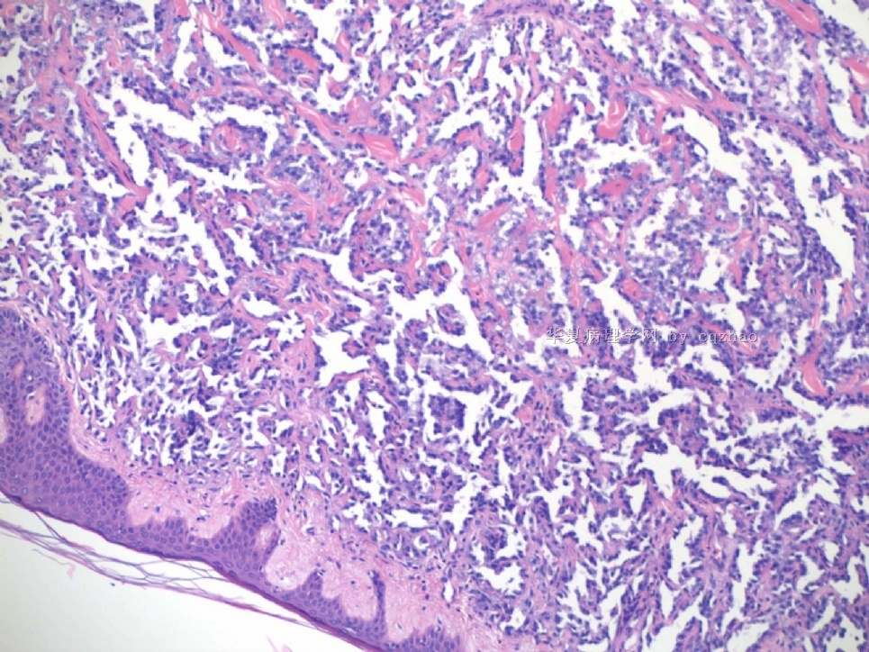
名称:图1
描述:图1
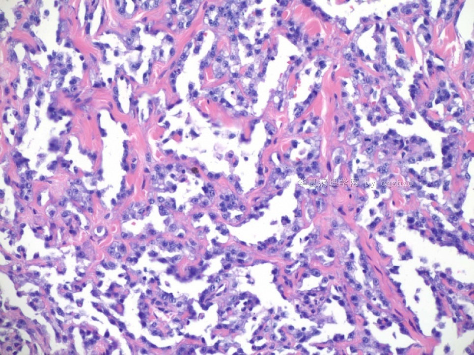
名称:图2
描述:图2
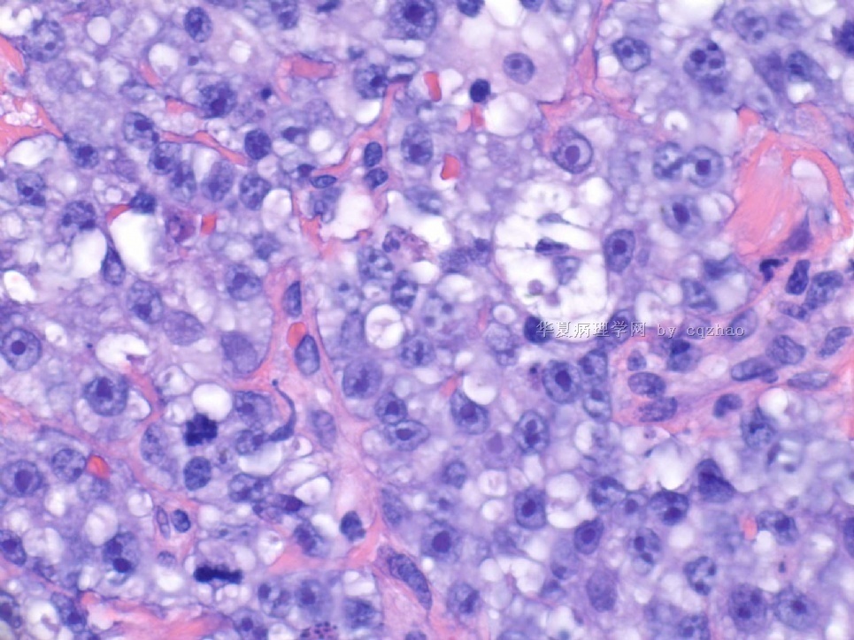
名称:图3
描述:图3
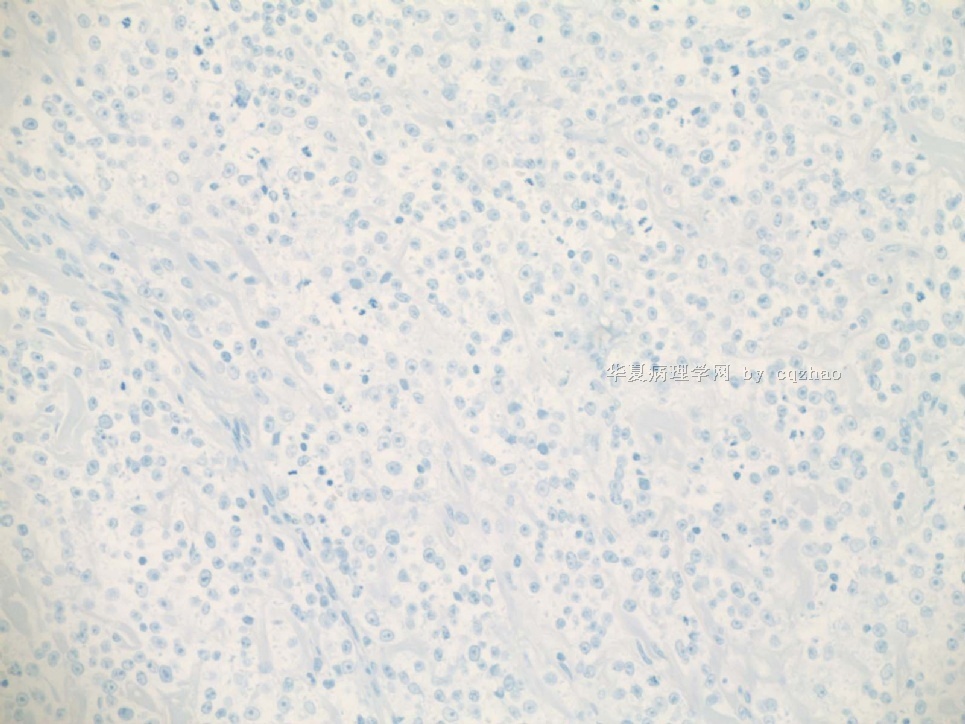
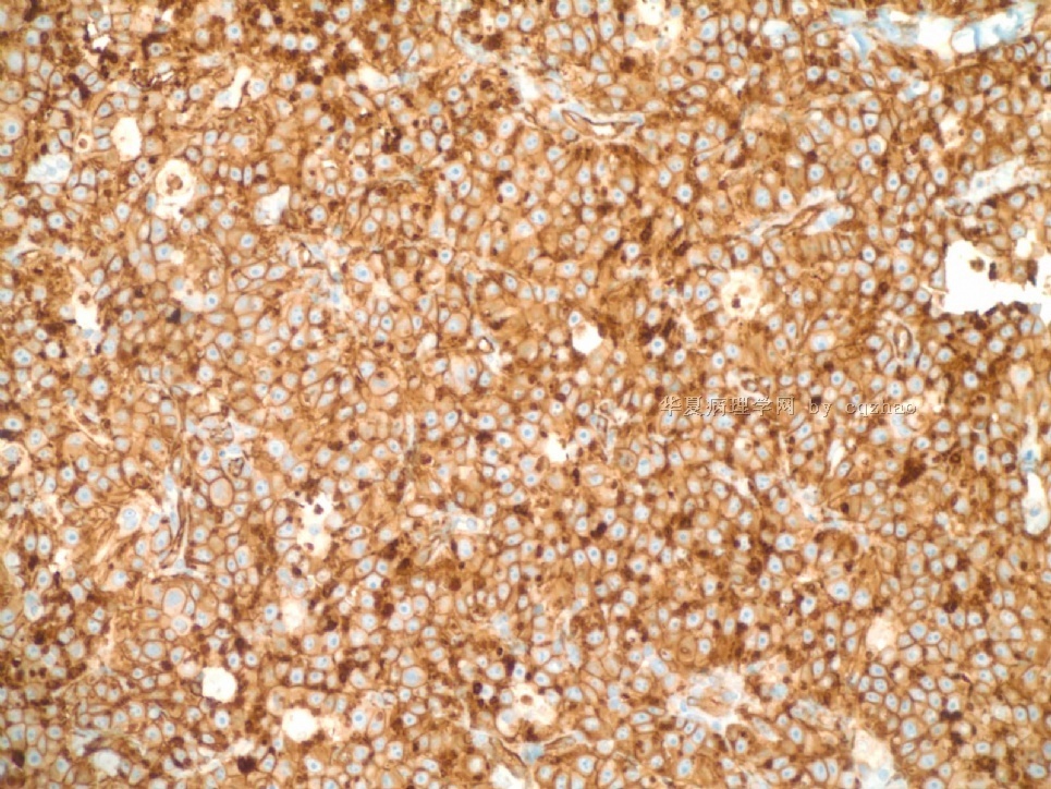
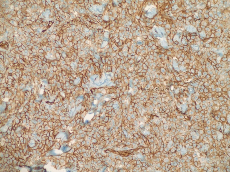
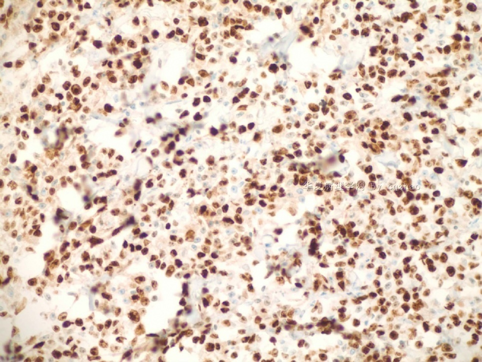
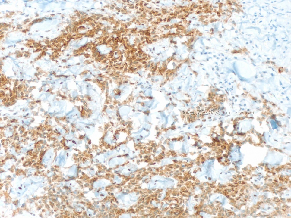
Just feel interested about ki67 proliferative index in high grade angiosarcoma. I did a ki67 stain for the purpose of education (not charge patient due to no meaning for dx). You can see the tumors are diffusely positive for ki67. I also asked the lab to do a D2-40 stain for education. We will not charge patients if the stains are for education. Also we will not report these results in the pathology reports.
D2-40 stain is positive for lymphatic endothelieum.
A few words for post radiation angiosarcona (AS)
1. The interval between radiation and AS is from 3 y to 12 y, mostly within 6 y.
2. Cutaneous presentation is more often than parenchynal AS.
3. Women with primary AS presentd with metastasis more often those with postradiaton AS.
4. Overall survival rates were no significantly different between primary and postradiation AS.
5. More cases are lymphatic origin in my impression. Correct me if it is wrong.
6. Histologic features of postradiation AS may be different from primary AS. You can find some papers to read for details if you are interested. I do not think it is very important for our dx. We need to consider the lesions and need some IHC.
Thank all of you for the discussion.
|
cqzhao老师回复: 因为对高级别血管肉瘤中ki-67指数感兴趣,所以出于研究的目的我们做了Ki-67的染色(因为没有诊断意义,所以不会向患者收钱的)。可以看到肿瘤组织广泛阳性。同时还让实验室做了一个D2-40的染色。如果我们是出于研究或学习的目的,那么这样的是不需要向患者收取费用的,当然我们在报告中也不会出现相关的结果。D2-40染色在淋巴管内皮细胞是阳性的、 |
|
关于放疗后血管肉瘤的几个关键词: |
1.血管肉瘤发病和放疗之间的间隔从3年到12年不等,大多是在6年内。
2.侵犯表皮的现象比原发的血管肉瘤更常见。
3.原发性血管肉瘤患者转移更常见。
4.原发性血管肉瘤和放疗后血管肉瘤患者总体累积生存率之间没有显著差异。
5.在我的印象中,大部分病例是来源于淋巴管上皮。(如果我说错了请纠正我一下)
6. 原发血管肉瘤和放疗后血管肉瘤组织学特征上是有差别的。如果你感兴趣,请多查点资料看一下。我认为这对于诊断其实并不重要。我们所要做的就是考虑到这个疾病然后上免疫组化确定。

- 赚点散碎银子养家,乐呵呵的穿衣吃饭
-
谢谢Dr.cqzhao,再次学习。一楼的图,细胞学特点应该是“上皮样细胞”,而不能一下子认定为“浆细胞样细胞”。因此它的鉴别诊断应该包括:癌、浆细胞瘤和上皮样血管肉瘤。
真正的乳腺浆细胞瘤:http://www.ipathology.org.cn/forum/forum_display.asp?keyno=271416
浆细胞瘤可能非常像癌:http://www.ipathology.org.cn/forum/forum_display.asp?keyno=140395

华夏病理/粉蓝医疗
为基层医院病理科提供全面解决方案,
努力让人人享有便捷准确可靠的病理诊断服务。
-
huisheng97 离线
- 帖子:263
- 粉蓝豆:22
- 经验:285
- 注册时间:2009-02-13
- 加关注 | 发消息
| 以下是引用cqzhao在2009-7-26 9:02:00的发言:
A few words for post radiation angiosarcona (AS) 1. The interval between radiation and AS is from 3 y to 12 y, mostly within 6 y. 2. Cutaneous presentation is more often than parenchynal AS. 3. Women with primary AS presentd with metastasis more often those with postradiaton AS. 4. Overall survival rates were no significantly different between primary and postradiation AS. 5. More cases are lymphatic origin in my impression. Correct me if it is wrong. 6. Histologic features of postradiation AS may be different from primary AS. You can find some papers to read for details if you are interested. I do not think it is very important for our dx. We need to consider the lesions and need some IHC. Thank all of you for the discussion. |
-
CHENYINQIAO 离线
- 帖子:547
- 粉蓝豆:0
- 经验:593
- 注册时间:2010-07-20
- 加关注 | 发消息
