| 图片: | |
|---|---|
| 名称: | |
| 描述: | |
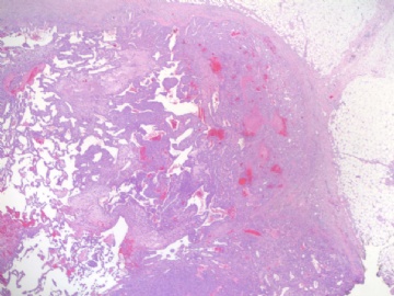
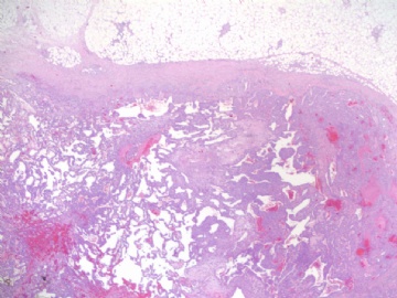
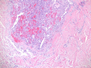
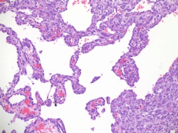
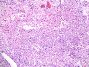
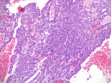
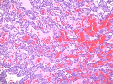
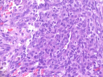
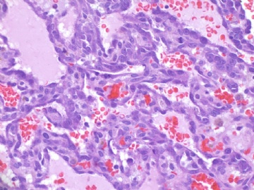
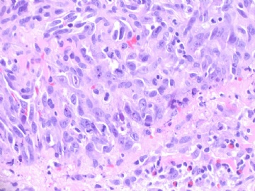
- B1840乳房高级别血管肉瘤(cqz-22)
| 姓 名: | ××× | 性别: | 年龄: | ||
| 标本名称: | |||||
| 简要病史: | |||||
| 肉眼检查: | |||||
About 70 y/f with breast lesion.
Noticed that 197 used the words "传说".I paste here another case for your guys. Do not say 传说 in future.
标签:乳腺高级别血管肉瘤
-
本帖最后由 于 2009-07-23 05:24:00 编辑
相关帖子
×参考诊断
