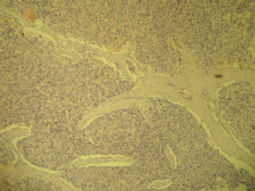
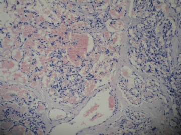
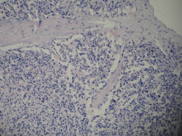
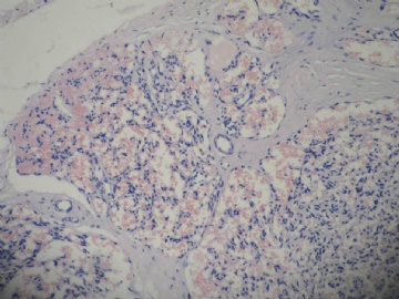
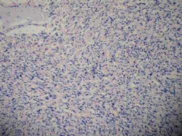
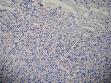
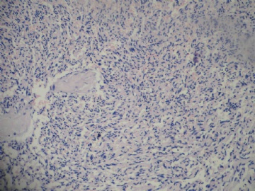
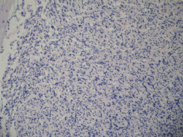
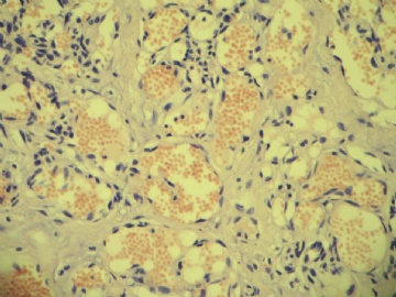
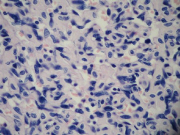
| 图片: | |
|---|---|
| 名称: | |
| 描述: | |
- B1838乳腺肿块
| 姓 名: | ××× | 性别: | 女 | 年龄: | 51 |
| 标本名称: | |||||
| 简要病史: | 右侧乳腺包块3-4个月 | ||||
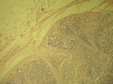
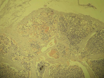
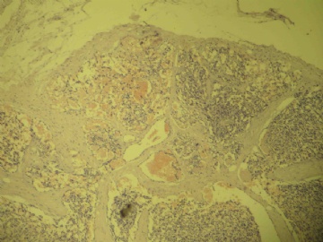
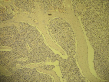
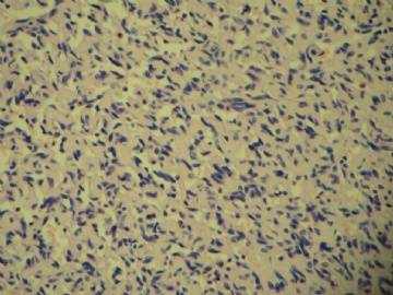
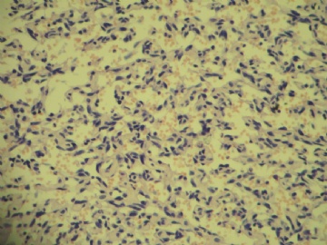
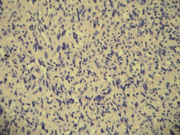
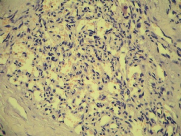
| 肉眼检查: | 肿块一枚,大小1*0.6*0.3cm,包膜完整,切面灰红,质中。 | ||||
相关帖子
- • 边界清楚的乳腺包块
- • 右乳肿块,新加免疫组化结果
- • 乳癌类型?
- • 乳腺肿块,请会诊
- • 左侧乳腺外上象限肿物,髓样癌?
- • 乳腺肿块
- • 女,29岁,发现右乳肿块,停止哺乳后手术切除肿块
- • 请求老师会诊,34岁女,乳腺肿物。
- • 乳腺癌类型?
- • 乳腺微创手术活检,诊断?
-
liguoxia71 离线
- 帖子:4174
- 粉蓝豆:3122
- 经验:4677
- 注册时间:2007-04-01
- 加关注 | 发消息
不会看,但看了些帖,收获不少。
1,乳腺为血管肉瘤好发部位这一,占乳腺肉瘤的9%,占乳腺肿瘤的0.03%。
2,乳腺血管肉瘤恶性度高,早期发生血行转移,为高度致死性肿瘤。
3,多发生于年青妇女,以40岁以下多,平均年龄34岁。
4,许多文献提到,几乎所有的乳腺血管性肿瘤均为恶性。换言之,在乳腺几乎没有良性血管瘤。此观点强调,当诊断良性血管瘤时,应注意防止漏诊。
5,血管肉瘤经常可见良性血管瘤区域或与皮肤血管瘤相似,故诊断良性血管瘤时,宜特别谨慎,必须注意多取材。
6,免疫组化示:vimentin(+)、CD31(+)、F8(+)、UEA-1(+)。
《肿瘤病理诊断与鉴别诊断学》P706。
It is not important for all of us to mention benign, atypical or malignant for these cases in the webs. The purpose for the web is to study. There are no experts or professors in the internet. Just people share their oppinion with others. Of cause you do not need to accept others' suggestion or concepts. Anyway in your daily practice you are the boss and you make your dx and sign your cases.
One thing I want to mention is that pathology diagnoses are not only benign and malignant, white and black. Why do we have so many borderline or atypical terms in many areas, for example atypical lobular/ductal hyperplasia, atypical papilloma in breast, serous/endoemtrioid/ mucinous borderline tumors in ovary? The reasons are we do not know exactly the clinical or biologic behavor for these kinds of cases. This is why people use the term of low malignant potential for these cases for some ovarian tumors. It is the same for these breast vascular lesions. We do not have a large study of these cases. We make the interpretation based on previous study results published. You are right to dx malignancy if these patients have poor prognosis in future. Also you are right to dx benign if these patients without mastectomy have no recurrent or metastic lesions in the future. If you are unlucky to have these cases in your practice you make your own judgment.
| 以下是引用cqzhao在2009-7-22 8:11:00的发言:
It is not important for all of us to mention benign, atypical or malignant for these cases in the webs. The purpose for the web is to study. There are no experts or professors in the internet. Just people share their oppinion with others. Of cause you do not need to accept others' suggestion or concepts. Anyway in your daily practice you are the boss and you make your dx and sign your cases. One thing I want to mention is that pathology diagnoses are not only benign and malignant, white and black. Why do we have so many borderline or atypical terms in many areas, for example atypical lobular/ductal hyperplasia, atypical papilloma in breast, serous/endoemtrioid/ mucinous borderline tumors in ovary? The reasons are we do not know exactly the clinical or biologic behavor for these kinds of cases. This is why people use the term of low malignant potential for these cases for some ovarian tumors. It is the same for these breast vascular lesions. We do not have a large study of these cases. We make the interpretation based on previous study results published. You are right to dx malignancy if these patients have poor prognosis in future. Also you are right to dx benign if these patients without mastectomy have no recurrent or metastic lesions in the future. If you are unlucky to have these cases in your practice you make your own judgment. |
abin译:在网上,我们谈论这些病例是良性、不典型或恶性并不重要。网上讨论的目的是学习。网上没有专家或教授,只是与别人分享观点。当然你们也不一定要接受别人的建议或观念。不管如何,在实际工作中,你自己是老板,你自己作出诊断并签发报告。
我要提到的一点,就是病理诊断不仅仅是良性和恶性,不是非黑即白。为什么在许多领域有很多交界性或不典型性病变的术语?例如乳腺的ALH、ADH、不典型乳头状瘤,卵巢的浆液性/内膜样/粘液性交界性病变。乳腺的血管病变也是如此。我们对此还没有大量研究。我们作出的解释是根据以前发表的研究结果。如果患者以后预后差,你诊断为恶性是合适的。当然如果患者不切除乳腺,将来不复发或不转移,你诊断为良性是对的。如果你运气不好,在实际工作中碰上这种病例,你需要自己判断。

- 努力的工作,快乐的生活!

