| 图片: | |
|---|---|
| 名称: | |
| 描述: | |
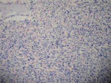
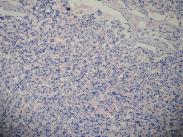
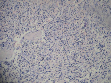
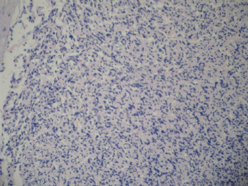
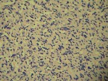
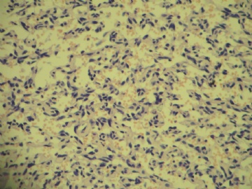
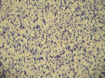
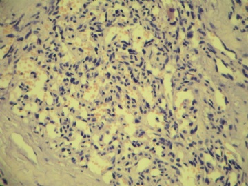
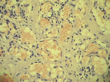
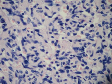
- B1838乳腺肿块
| 姓 名: | ××× | 性别: | 女 | 年龄: | 51 |
| 标本名称: | |||||
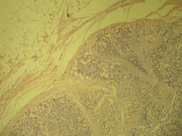
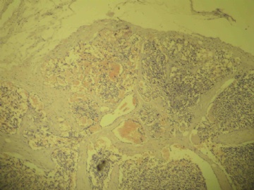
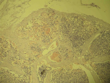
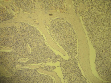
| 简要病史: | 右侧乳腺包块3-4个月 | ||||
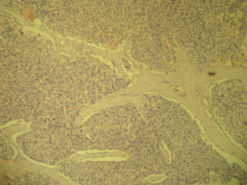
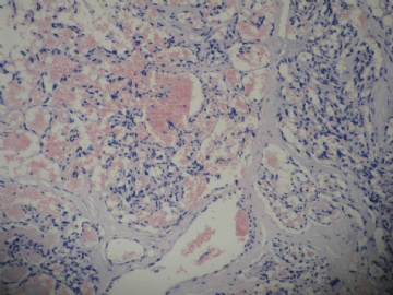
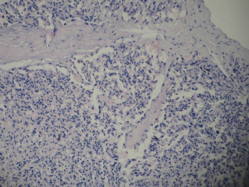
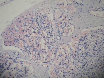
| 肉眼检查: | 肿块一枚,大小1*0.6*0.3cm,包膜完整,切面灰红,质中。 | ||||
相关帖子
- • 边界清楚的乳腺包块
- • 右乳肿块,新加免疫组化结果
- • 乳癌类型?
- • 乳腺肿块,请会诊
- • 左侧乳腺外上象限肿物,髓样癌?
- • 乳腺肿块
- • 女,29岁,发现右乳肿块,停止哺乳后手术切除肿块
- • 请求老师会诊,34岁女,乳腺肿物。
- • 乳腺癌类型?
- • 乳腺微创手术活检,诊断?
-
jianshu322 离线
- 帖子:447
- 粉蓝豆:17
- 经验:1405
- 注册时间:2008-12-22
- 加关注 | 发消息
I favor a dx of atypical hemagioma. Patient should have completely excision biopsy. Clinical-imaging follow-up. The reasons are similar to the case cqz-20. I have a lot of discussion in this link.
http://www.ipathology.cn/forum/forum_display.asp?keyno=158702
-
Quickly review the discussion above. I noticed some pathologists with excellent analysis of these kinds of cases. However, I feel a little disappointed for some pathologists' direct diagnosis. I might spend a lot of time and have a little effect on many pahtologists.
最近有时会想一个问题,美国的朋友打电话有时也討论此问题: 在美国工作紧张,压力大, 是否有价值,有效果,花费许多时间和精力在华夏网站上。
Difficult to type Chinese.
Many people here just saw the photos and then gave a dx. Just wonder what you can get in this way.
In fact there are too many expert pathologists in China. They have more responsibility to teach young pathologists in the local hospitals. They have the responsibility to develop the pathology in China. It is easy for them to do these issues. I know there are relative numbers of American Chinese pathologists whoactively paticipate the discussion and lectures in this web. Many Chinese expert pathologists gave lecture in this web. However, how many Chinese expert pathologists attend actively the case discussion? Some chinese pathologists told me that Chinese expert pathologists or professors are very busy for daily work, for book, for meeting talk. Seem that we in the US are not busy. 无语.
-
本帖最后由 于 2009-07-21 18:59:00 编辑
对不起Dr. zhao,我首先接受您的批评。首先我要检讨,我做的就不好。

我想,可能是中国文化造成的这种现象。您知道,在国内的课堂上很少有人向老师提问题。在国内讲课也很少有人与教授讨论问题。并不是我们的学习积极性不够高。我个人就是个例子:“敏于行而讷于言”,总感觉不如别人,我所知道的可能别人都知道了,别人知道的我却不知道。
国内同行看贴的多,回帖的少,可能有下列情况:一是看不懂不敢说;二是拿不准不好意思说,怕说错;三是都想听听别人怎么说,看看别人的意见,只是喜欢向别人学习。再说国内大多数基层医院,病理医生手底下可以参考的资料实在是太少了,很难参加讨论。
以后一定改正!
也希望广大网友多多发表意见。

- 博学之,审问之,慎思之,明辨之,笃行之。
| 以下是引用cqzhao在2009-7-18 9:27:00的发言: Quickly review the discussion above. I noticed some pathologists with excellent analysis of these kinds of cases. However, I feel a little disappointed for some pathologists' direct diagnosis. I might spend a lot of time and have a little effect on many pahtologists. |
| 以下是引用cqzhao在2009-7-18 20:00:00的发言:
最近有时会想一个问题,美国的朋友打电话有时也討论此问题: 在美国工作紧张,压力大, 是否有价值,有效果,花费许多时间和精力在华夏网站上。 Difficult to type Chinese. Many people here just saw the photos and then gave a dx. Just wonder what you can get in this way. In fact there are too many expert pathologists in China. They have more responsibility to teach young pathologists in the local hospitals. They have the responsibility to develop the pathology in China. It is easy for them to do these issues. I know there are relative numbers of American Chinese pathologists whoactively paticipate the discussion and lectures in this web. Many Chinese expert pathologists gave lecture in this web. However, how many Chinese expert pathologists attend actively the case discussion? Some chinese pathologists told me that Chinese expert pathologists or professors are very busy for daily work, for book, for meeting talk. Seem that we in the US are not busy. 无语. IN fact we are very interested in your discussions and have make much progress both in English and speciality hereby we feel very thankful for my honoured fellows as well as my best teachers abroad.We are not purposely give direct diagnosis because of different kind of reasons including time and speciality level.but for this case,because of its lobular structures i insist on capillary angioma though most of the case of vascular origine in breast are angiosarcoma.Finally please forgive me my shabby English! |

朱正龙

