| 图片: | |
|---|---|
| 名称: | |
| 描述: | |
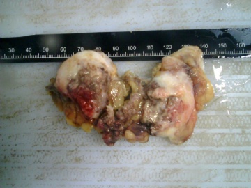
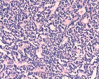
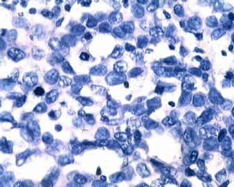
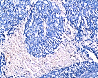
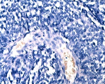
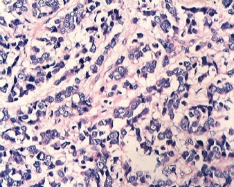
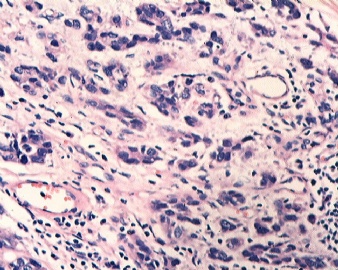
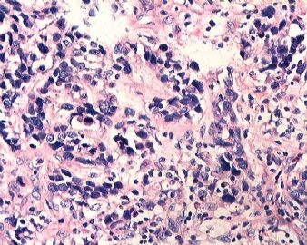
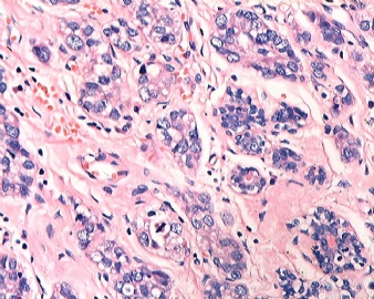
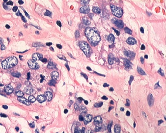
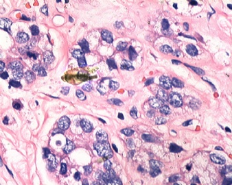
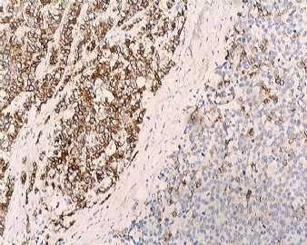
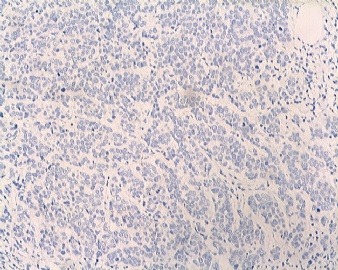
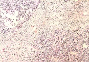
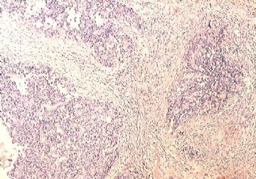
- B1833乳腺肿瘤,请教什么类型
| 姓 名: | ××× | 性别: | 女 | 年龄: | 46 |
| 标本名称: | 乳腺肿块 | ||||
| 简要病史: | 发现肿物3天 | ||||
| 肉眼检查: | |||||
-
本帖最后由 于 2009-06-13 13:40:00 编辑
相关帖子
Thank wq_9603 for your excellent translation.
The % of positivity of breast tumor for GCDFP depends on the tumor types you choose. Most breast ca with apocrine features are positive for GCDFP. In fact mamoglobin is more sensitive for breast carcinoma. Of cause endometrial carcinomas also are positive for mamoglobin.
-
关于GCDFP-15:中文译名大囊肿性疾病液体蛋白-15,是乳腺囊肿液中的一种组成蛋白,可在任何具有大汗腺特征的细胞中表达。除了乳腺之外,可在涎腺和汗腺的腺泡结构中表达,也可在皮肤、外阴Paget病和前列腺中表达。有著作提出:62%-77%的乳腺癌、以及涎腺和皮肤附属器的肿瘤表达GCDFP-15,其阳性表达对诊断乳腺癌有98%-99%的特异性。比如在同一本著作中提到:在鉴别乳腺与卵巢恶性肿瘤时候,如果GCDFP-15阳性则一般认为是乳腺原发,但是阴性表达则不能提供诊断帮助。

- 赚点散碎银子养家,乐呵呵的穿衣吃饭
-
漫游人老师意见:
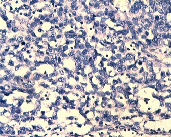
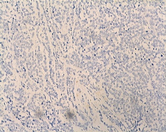
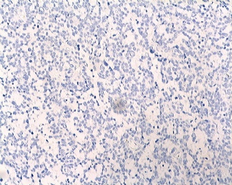
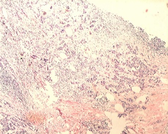
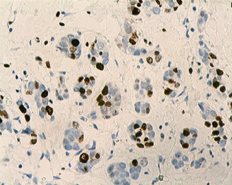
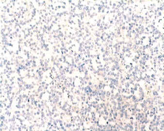
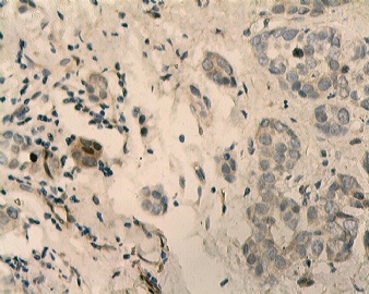
免疫组化GCDFP-15染色对确定乳腺原发很有帮助。我注意到在最后几张HE图片中有很多炎症细胞浸润,特别是在低分化的区域(您指的是倒数第二、三张吗?)。少数细胞PR阳性,是不是残留的正常乳腺上皮呢?如果我的猜测是对的,那么上述三个阴性结果(ER、PR、HER2)表明此病理可能是基底细胞样癌。尽管形态学上不太典型,但是在作出最后诊断之前还是最好排除其他可能性。正如abin所分析的,还需要多做点标记。我会建议排除一些神经内分泌方面分化(可能在切片上比我们看图片更容易做出判断),其他来源的标记物也要做一下,如TTP-1,CK20,CK7,尽管临床信息方面没有这方面提示。
cqzhao老师意见:
同意以上分析,但是要注意到大部分的乳腺癌病例GCDFP是阴性的。至于基底细胞样癌的话,建议加做CK5(或CK5/6)、CK14、CK17、EGFR

- 赚点散碎银子养家,乐呵呵的穿衣吃饭
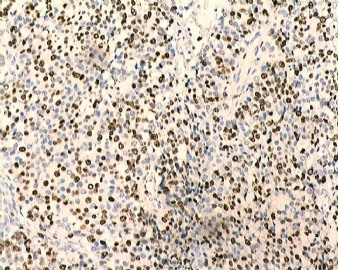
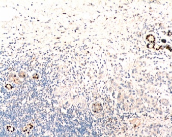
GCDFP-15 stain may be of help to confirm a breast primary.
I noticed that the latter H&E photos showed more inflammatory cell infiltrate especially in the poorly differentiated areas.
I'm afraid the occasional positive PR staining cells are from residual normal breast acini. If this is correct, triple negative will put this case into a basal type carcinoma.
Since the morphology is atypical, it's better to exclude other possibilities before giving final decision.
Like abin suggested, further markers for basal type. I would suggest again to exclude neuroendocrine differentiation (Maybe you see better on glass slide than pictures). Markers indicating other primaries can also be tried, such as TTP1, Ck20, CK7, although clinically not indicated.
-
本帖最后由 于 2009-06-17 10:44:00 编辑
add to
 |
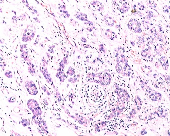
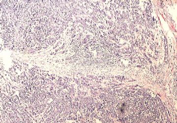
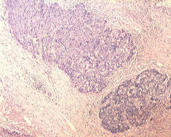
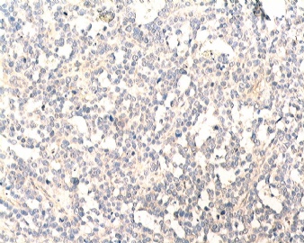
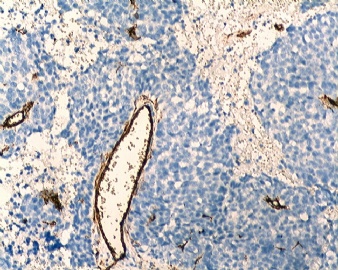
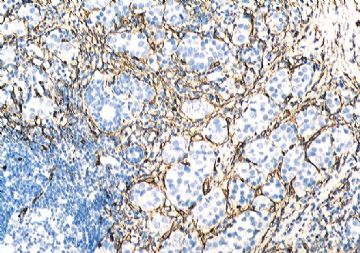
Based on IHC and low power photos
1. Mostly one tumor with two growth patterns
2. More like carcinoma, especially adenocarcarcinoma
3. Primary or secondary. Favor primary: no other organ lesions. Common things are common. Of cause most breast tumors are primary. However it is not a classic breast tumor. Did the pt have x-ray exam for most organs?
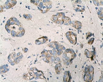
4. If it is a primary breast carcinoma the next question is what types. Ductal vs lobular. Favor ductal ca due to the glandular formation and positive E-cad (membrane stain).
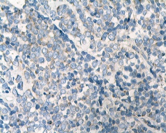
5. ER, PR stains: If they are positive the tumor is from breast or gynecologic system. PR seems weakly positive in rare cells (not sure).
It will be more easy to read slides
Thank Quan zi for the challenge case. Wish more people share with us your oppinion. We still do not know the final dx yet.
cz
基于免疫组化和低倍镜下的图片
1.主要表现是一种含有两种生长模式的肿瘤
2.更大的可能是癌,特别是腺癌
3.原发或是继发。倾向于原发:无其它部位的病变。通常先考虑常见的,当然大多数乳腺肿瘤都是原发的。然而这不是一类经典的乳腺肿瘤,这位病人有做过其它器官的X线检查吗?
4.如果此例是原发性的乳腺癌,那么接下来的问题就是它是属于哪一型的呢,导管来源或小叶来源?倾向于导管癌因为有腺腔形成并且E-cad表达阳性。
5.ER,PR染色:如果此肿瘤是来自于乳腺或女性生殖系统,那么ER,PR染色应当为阳性。
PR染色在少许细胞可见到弱阳性表达(不是很确定)
如果是看切片将会更容易些。
谢谢全子提供如此有挑战性的病例,希望更多的人和我们一起分享你们的意见,
我们仍然不知道最终结果。from cz
海棠依旧翻译
Thank 海棠's excellent translation. cz
-
jixiangrui 离线
- 帖子:17
- 粉蓝豆:1
- 经验:17
- 注册时间:2009-05-18
- 加关注 | 发消息
-
本帖最后由 于 2009-06-16 11:40:00 编辑
| 以下是引用cqzhao在2009-6-15 22:39:00的发言:
Quan zi: please show us some low power phtos. I want to know the relation of the first 6 photos to others. Did you use different stains? Why did two groups of photos show different color? |
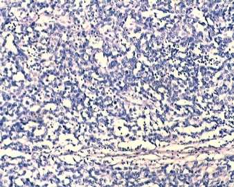
低倍图像
不同颜色是因为我的图像采集器不稳定
嘿嘿

