| 图片: | |
|---|---|
| 名称: | |
| 描述: | |
- 左颞叶肿瘤
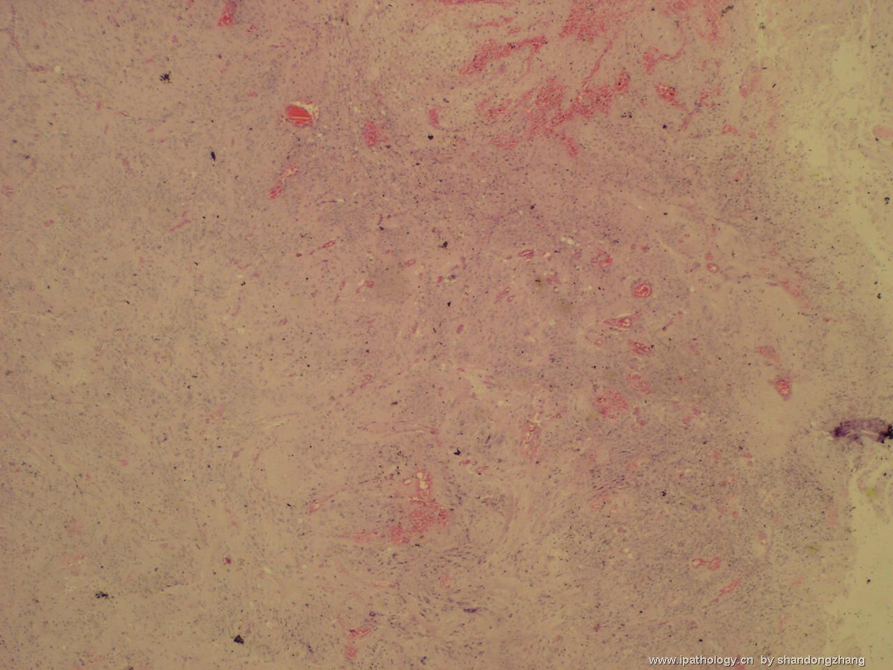
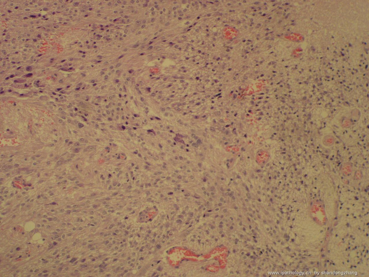
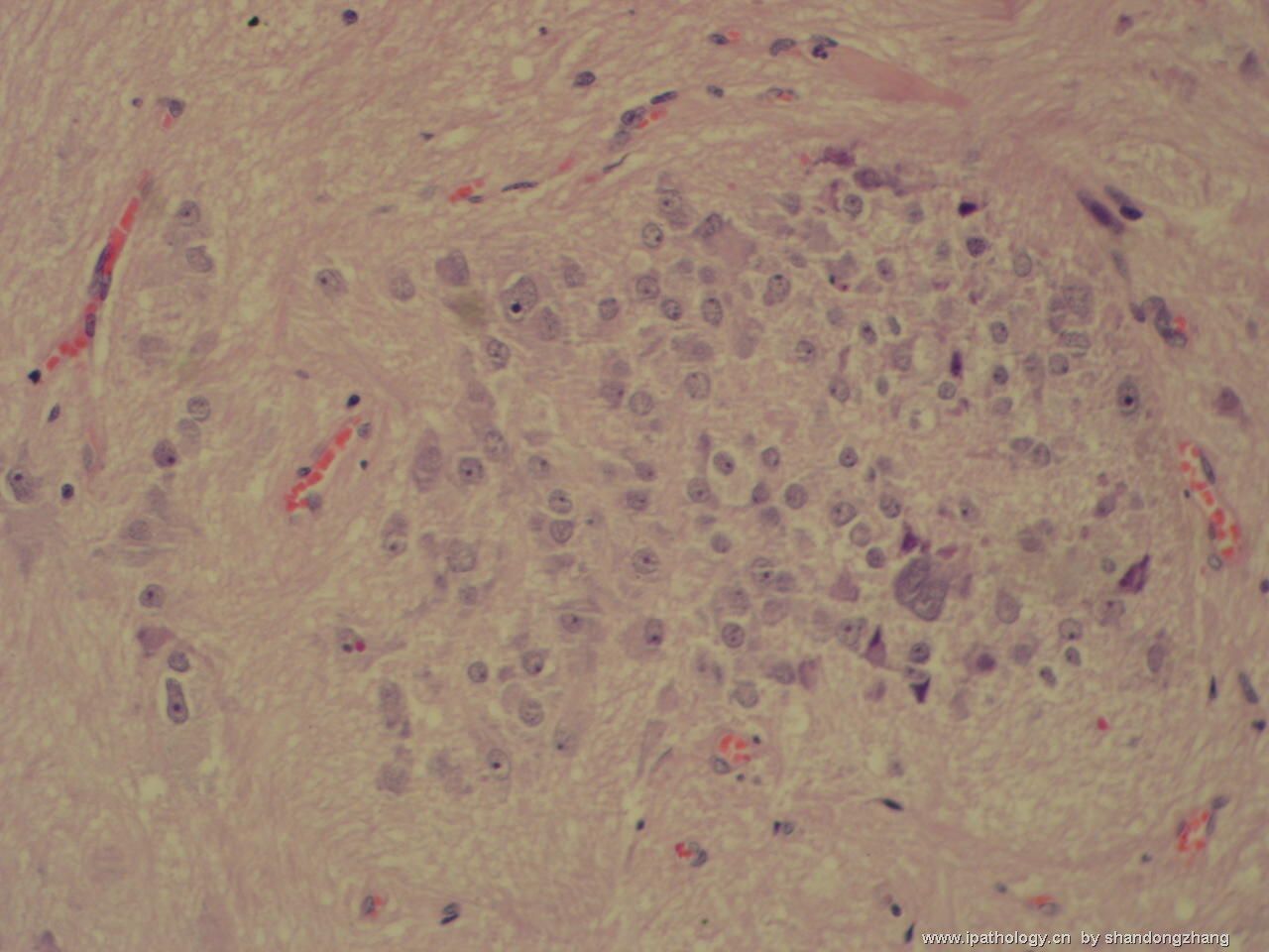
First and importantly, this case is not 发育不良性小脑节细胞瘤(WHO grade I dysplastic cerebellar gangliocytoma or Lhermitte-Duclos disease) that has very specific gross and histopathology, and is sometimes associated with Cowden disease. Figure 1 shows possible necrosis in the congested corner, but this is not associated with features of a high grade neoplasm. Grade III anaplastic ganglioglioma, grade IV glioblastoma arising from a ganglioglioma, and grade IV ganglioneuroblastoma are not difficult to diagnose.
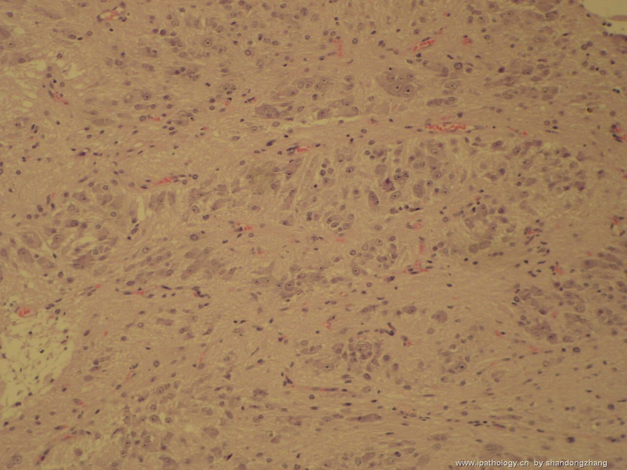
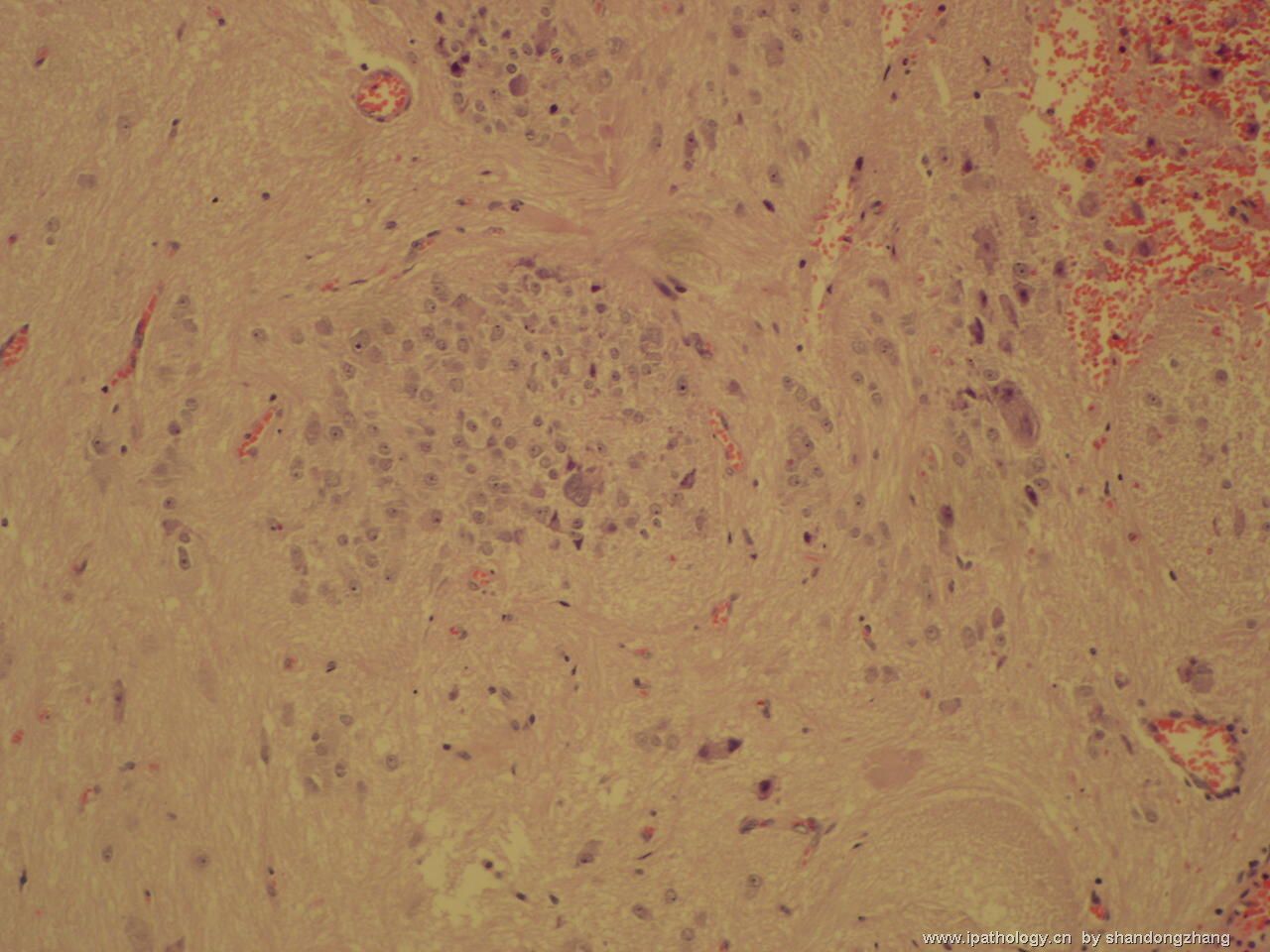
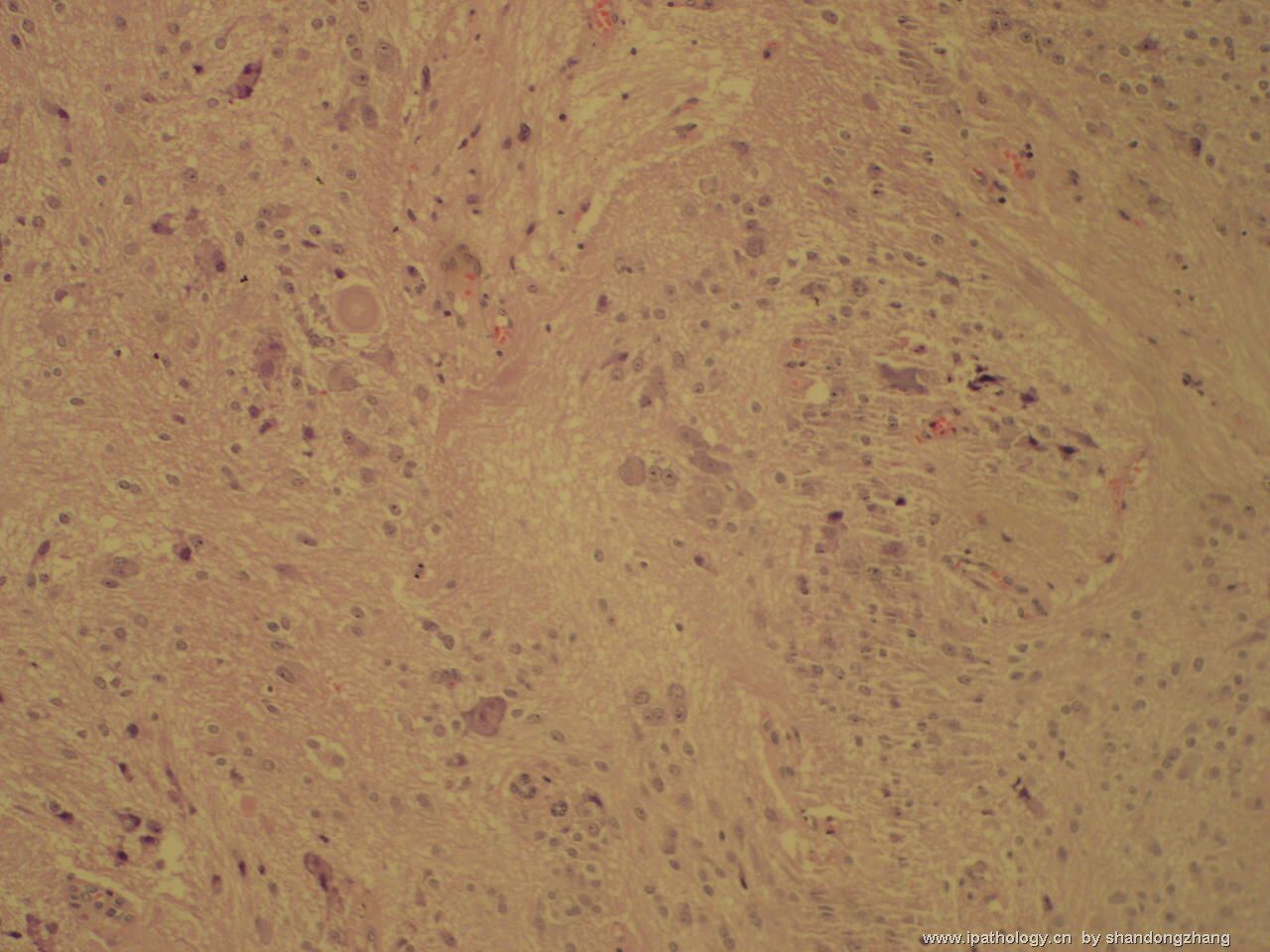
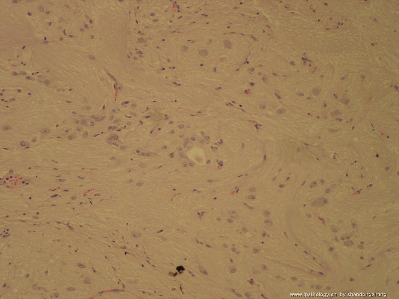
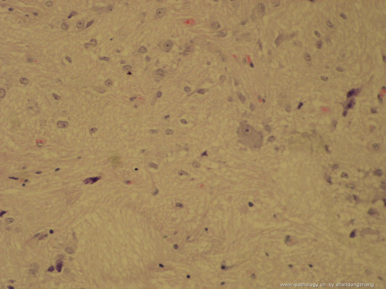
Distinction between grade I gangliocytoma and grade I ganglioglioma is probably not an important one, since they have similar if not identical biologic behavior and prognosis. Certainly, one has to see a gliomatous component to call a neuronal tumor a ganglioglioma. This judgment can be very subjective - "all in the eyes of the beholder". From the photos you kindly provided, I have not seen a definite gliomatous component in this case. Short of seeing relatively large areas (greater than one 100x microscopic field) of glial neoplasm without admixed dysplastic neurons, I usually would not call it a ganglioglioma. This is my personal criterion and by no means scientific. Naturally, I cannot be absolutely certain about this point without examining the histologic slides myself.
The distinction between WHO grade I and grade II gangliogliomas is not as clear cut as it seems. If one uses the conventional definitions, grade I ganglioglioma is very uncommon. Remember, pure gangliocytomas often contain eosinophilic granular bodies. Therefore, one would like to see other characteristic features of pilocytic astrocytoma (biphasic growth pattern, mucinous microcysts, Rosenthal fibers, uniform piloid cells) before calling any tumor grade I ganglioglioma. In the photos uploaded so far, I have not seen these features. Finally, when the gliomatous component is very bland and I cannot decide, I would just diagnose it as a grade I~II ganglioglioma. I hope my comment is more enlightening than confusing.

聞道有先後,術業有專攻
| 以下是引用mjma 在2006-10-7 13:24:00的发言: Though Figure No 5 is cellular, most cells in this neoplasm have cytologic features of ganglion cells or neurons. Calcification is seen as well. That make this a WHO grade I gangliocytoma. I do not see a gliaoma component to make this a ganglioglioma. In general, WHO grade I gangliocytomas are benign lesions found mainly during workup of seizure disorders. Grading of a ganglioglioma depends on the co-existing glioma (usually astrocytoma) present. It could be grade I (if the glioma is a pilocytic astrocytoma), grade II (diffuse or fibrillary astrocytoma), grade III (anaplastic astrocytoma) or, very rarely, grade IV (glioblastoma). Sometimes, differentiated ganglion cells are found with primitive embryonal cells that also express neuronal markers (NeuN, NSE. synaptophysin, neurofilaments). That combination qualifies the lesion as a WHO grade IV ganglioneuroblastoma. |
-
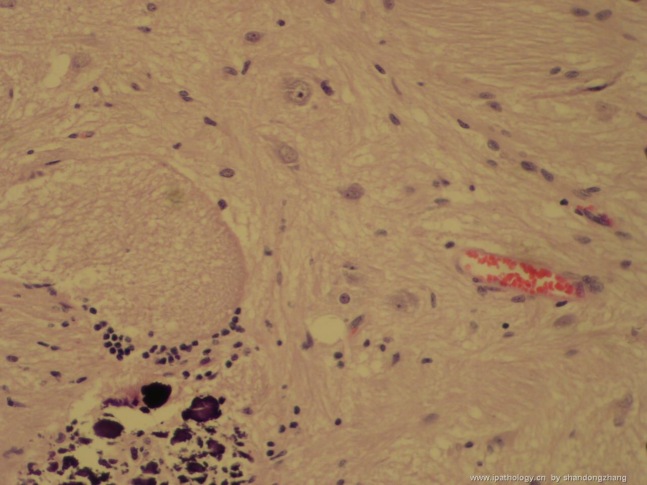
Though Figure No 5 is cellular, most cells in this neoplasm have cytologic features of ganglion cells or neurons. Calcification is seen as well. That make this a WHO grade I gangliocytoma. I do not see a gliaoma component to make this a ganglioglioma. In general, WHO grade I gangliocytomas are benign lesions found mainly during workup of seizure disorders. Grading of a ganglioglioma depends on the co-existing glioma (usually astrocytoma) present. It could be grade I (if the glioma is a pilocytic astrocytoma), grade II (diffuse or fibrillary astrocytoma), grade III (anaplastic astrocytoma) or, very rarely, grade IV (glioblastoma). Sometimes, differentiated ganglion cells are found with primitive embryonal cells that also express neuronal markers (NeuN, NSE. synaptophysin, neurofilaments). That combination qualifies the lesion as a WHO grade IV ganglioneuroblastoma.

聞道有先後,術業有專攻

