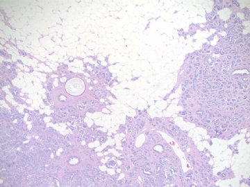
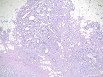
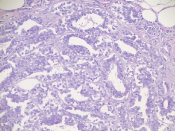
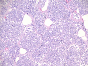
| 图片: | |
|---|---|
| 名称: | |
| 描述: | |
- B1813乳房肿块, 少见,但easy to make dx (czq-19)
-
dingdingxiao 离线
- 帖子:1
- 粉蓝豆:1
- 经验:1
- 注册时间:2009-06-23
- 加关注 | 发消息
cqzhao老师的回复:
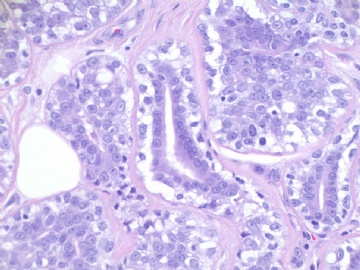
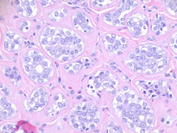
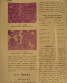
谢谢天山望月的分析。这是AFIP的病例,年龄50岁,肿瘤直径1.5cm。免疫组化显示肌上皮细胞增生。
简单浏览了一下大家的分析,几乎都回答正确。这是一个腺肌上皮病变(adenomyoepithelial lesion),两种细胞成分。乳腺的腺肌上皮瘤(Adenomyoepithelioma ,AME)比较少见,其特征就是上皮和肌上皮共存。第一次完全的阐述是由Hamperl于1970年发表的。
哪位能分别总结一下良性AME和恶性AME的特征?我没有查阅资料,所以对这方面的界定不是很确定了。希望有人查一下与大家分享,谢谢
我们的网站上有很多的病例。仅仅是看一下提供的图片然后“猜”一个诊断是没有意义的。典型的图像在书上或者光碟上面是很容易看到的。上网的好处就在于需要你思考一下鉴别诊断,总结一下病例,讨论一下你的观点,与大家分享……。我们都应该是参与者而不仅仅是阅览者……

- 赚点散碎银子养家,乐呵呵的穿衣吃饭
There are many cases in this website. It is meaningless if we see the photos and give a guess diagnosis. It is easy to have many classic pictures for all lesions in the text books, CD et al.
The advantage here online is that you can think over the differential dx, summary the cases, discuss the cases and share your oppinion with all others. We all are participants, not only readers.
Quickly review above interpretation from all of you. It is good almost all of you got the correct point. Clearly it is a adenomyoepithelial lesion, two cell component.
Adenomyoepithelioma (AME) of the breast is an uncommon tumor characterized by the presence of both epithelial and myoepithelial cells; its first full description was published in 1970 by Hamperl.
Could some one sumarry the features of benign and maligant AME? I have not check books or literatures yet. So In fact I am not clear about the definition now. Hope some one will do the search and share with us here. Thanks, cz
仔细阅读图片,组织结构:肿瘤无包膜,浸润性生长,呈巢状或管状排列,部分腔面有一层腺上皮,部分为均一细胞,大小一致,胞浆透亮,核染色质细腻,核仁明显,间质纤维化玻璃样变。
考虑:
1、腺肌上皮瘤/病?
2、腺肌上皮癌?(阿克曼病理学中描述:所有透明细胞型的腺肌上皮瘤都具有恶性潜能。第九版,890页)
3、微腺腺病:在脂肪中浸润性生长,但为单层细胞,此例不支持。
乳腺的肌上皮病变与涎腺的相似,良性瘤,体积小,多在1cm下,边界清,无浸润性生长,细胞无异型性。
恶性肌上皮i瘤,体积大,多结节浸润性生长,细胞异型性可小到明显异型。
此例多结节状浸润性生长,透明肌上皮细胞,考虑低度恶性可能性大,不知患者年龄、肿瘤大小等临床情况如何?IHC标记:CK5、SMA、S-100、GFAP、Calponin、P63、Ki67、Ⅳ胶原等情况如何?
仅为个人观点,请Dr.zhao详细讲解!谢谢!

- 广州金域病理
