共4页/62条首页上一页1234下一页尾页
回复:62 阅读:7275
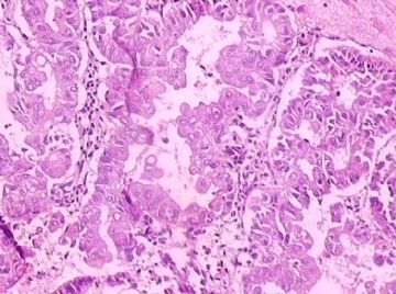
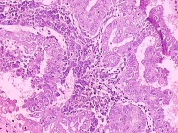
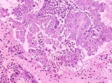
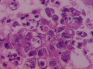
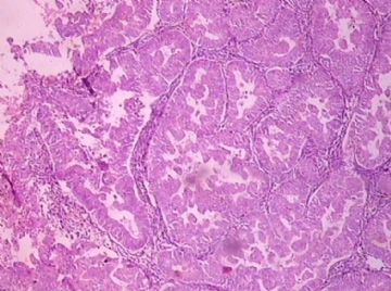
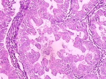
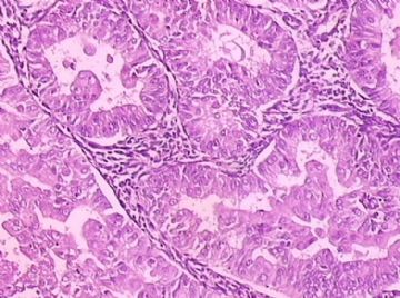
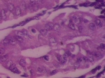
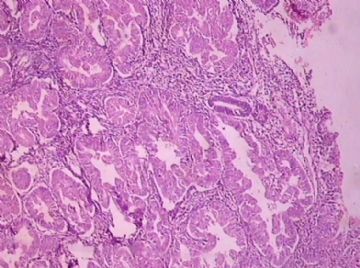
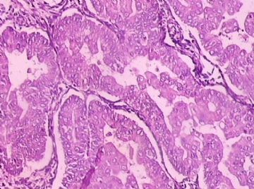
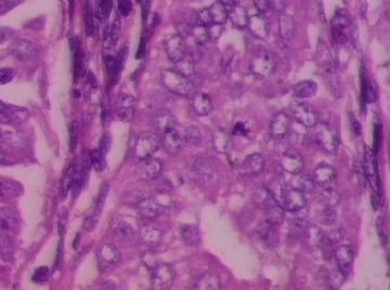
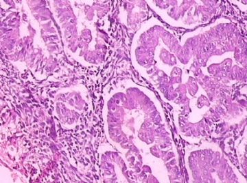
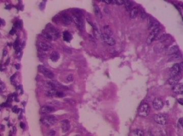
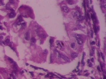
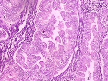
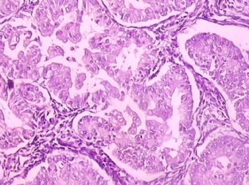
B172344岁子宫内膜标本,请各位老师会诊,谢谢

千百合 离线

帖子:1294
粉蓝豆:121
经验:1764
注册时间:2006-12-13
加关注  |  发消息
楼主 发表于 2009-05-14 20:35|举报|关注(0)
浏览排序[ 顺序 逆序 楼主 支持 精彩 ]  快捷回复
姓    名: ××× 性别:  女 年龄:  44
标本名称: 刮宫标本
简要病史:  阴道不规则流血20天
肉眼检查:  灰红组织一堆,直径3厘米
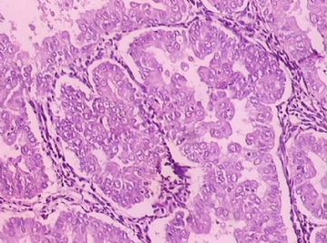
  • 44岁子宫内膜标本,请各位老师会诊,谢谢图1
    图1
  • 44岁子宫内膜标本,请各位老师会诊,谢谢图2
    图2
  • 44岁子宫内膜标本,请各位老师会诊,谢谢图3
    图3
  • 44岁子宫内膜标本,请各位老师会诊,谢谢图4
    图4
  • 44岁子宫内膜标本,请各位老师会诊,谢谢图5
    图5
  • 44岁子宫内膜标本,请各位老师会诊,谢谢图6
    图6
  • 44岁子宫内膜标本,请各位老师会诊,谢谢图7
    图7
  • 44岁子宫内膜标本,请各位老师会诊,谢谢图8
    图8
  • 44岁子宫内膜标本,请各位老师会诊,谢谢图9
    图9
  • 44岁子宫内膜标本,请各位老师会诊,谢谢图10
    图10
  • 44岁子宫内膜标本,请各位老师会诊,谢谢图11
    图11
  • 44岁子宫内膜标本,请各位老师会诊,谢谢图12
    图12
  • 44岁子宫内膜标本,请各位老师会诊,谢谢图13
    图13
  • 44岁子宫内膜标本,请各位老师会诊,谢谢图14
    图14
  • 44岁子宫内膜标本,请各位老师会诊,谢谢图15
    图15
  • 44岁子宫内膜标本,请各位老师会诊,谢谢图16
    图16
  • 44岁子宫内膜标本,请各位老师会诊,谢谢图17
    图17
标签:子宫内膜 增生 局灶癌变
本帖最后由 于 2009-05-14 20:36:00 编辑
0
signature
努力的工作,快乐的生活!
×参考诊断
复杂性不典型增生,局灶癌变

闹闹 离线

帖子:944
粉蓝豆:24
经验:1921
注册时间:2007-07-25
加关注  |  发消息
41 楼    发表于2009-05-14 20:54:00举报|引用
返回顶部 | 快捷回复
 复杂型增生伴不典型增生
0
回复

宁静致远 离线

帖子:4242
粉蓝豆:2853
经验:6556
注册时间:2007-03-27
加关注  |  发消息
42 楼    发表于2009-05-14 21:29:00举报|引用
返回顶部 | 快捷回复
 高分化子宫内膜样腺癌
0
回复

njwbhuang 离线

帖子:712
粉蓝豆:2265
经验:1404
注册时间:2007-11-23
加关注  |  发消息
43 楼    发表于2009-05-14 21:35:00举报|引用
返回顶部 | 快捷回复
 

Uterine Endometrioid Carcinoma with Small Nonvillous Papillae: An Analysis of 26 Cases of a Favorable-Prognosis Tumor To Be Distinguished from Serous Carcinoma.

James Homer Wright Pathology Laboratories of the Massachusetts General Hospital and the Department of Pathology, Harvard Medical School.

We have encountered a number of endometrioid carcinomas with small papillary buds lacking fibrovascular cores that could be confused with the small cellular papillae of serous papillary carcinoma (SPC). We have designated these tumors "endometrioid carcinoma with small nonvillous papillae" (ECSP). Because they have not been investigated previously we analyzed 26 examples and compared their features with those of 21 SPCs of the uterus. Three hundred and ninety consecutive cases of endometrial carcinoma diagnosed between January, 1989, and January, 1994, were retrieved from our hospital files; 26 (6.7%) of them, (8% of the endometrioid carcinomas) were identified as ECSP, and 21 (5.4%) as SPC. Tumors were classified as ECSP when the small papillae were present within the glands of otherwise typical endometrioid carcinoma or on the villous projections of villoglandular endometrioid carcinoma. Most of the papillae were in the form of buds of cells with ample eosinophilic cytoplasm and a low nuclear-to-cytoplasmic ratio, but some papillae had a more complex pattern. The papillae arose on a background of International Federation of Gynecology and Obstetrics (FIGO) grade 1 or 2 endometrioid carcinomas, which showed squamous differentiation in half the cases. SPCs were identified according to generally accepted criteria. The mean age of the patients with ECSP was 67 years, intermediate between that of the patients with endometrioid carcinoma lacking small nonvillous papillae (62 years) and that of the patients with SPC (71 years). Patients with ECSP more frequently presented at an earlier stage (73% stage I/II) than those with SPC (29% stage I/II). The overall 5-year survival of patients with ECSP was 84% (95%CI: 0.68-1), more than double that of patients with SPC, 33% (95%CI: 0.10-0.56). ECSP may be confused with SPC on microscopic examination but has clinical and pathological features similar to those of endometrioid carcinoma lacking small nonvillous papillae, and unlike SPC, should be treated in the same manner as the former. Int J Surg Pathol 8(4):279-289, 2000

0
回复

wangdingding 离线

帖子:1474
粉蓝豆:98
经验:6042
注册时间:2006-10-19
加关注  |  发消息
44 楼    发表于2009-05-14 21:38:00举报|引用
返回顶部 | 快捷回复
 间质似有纤维化,不能排除癌。
0
回复

甲乙丁 离线

帖子:70
粉蓝豆:547
经验:75
注册时间:2009-03-20
加关注  |  发消息
45 楼    发表于2009-05-14 21:50:00举报|引用
返回顶部 | 快捷回复
 支持楼主的意见.
0
回复

qxrmyyljw 离线

帖子:240
粉蓝豆:24
经验:254
注册时间:2008-11-20
加关注  |  发消息
46 楼    发表于2009-05-14 22:04:00举报|引用
返回顶部 | 快捷回复
 复杂型增生伴不典型增生
0
回复
signature
知足常乐

文长江 离线

帖子:1042
粉蓝豆:139
经验:1438
注册时间:2007-03-21
加关注  |  发消息
47 楼    发表于2009-05-14 22:13:00举报|引用
返回顶部 | 快捷回复
以下是引用宁静致远在2009-5-14 21:29:00的发言:

 高分化子宫内膜样腺癌

0
回复

abin 离线

帖子:7416
粉蓝豆:6014
经验:11787
注册时间:2006-10-08
加关注  |  发消息
48 楼    发表于2009-05-14 22:22:00举报|引用
返回顶部 | 快捷回复
 同意njwbhuang的意见,可以直接报子宫内膜腺癌,可能是ECSP。
0
回复
signature

华夏病理/粉蓝医疗

为基层医院病理科提供全面解决方案,

努力让人人享有便捷准确可靠的病理诊断服务。


liguoxia71 离线

帖子:4174
粉蓝豆:3122
经验:4677
注册时间:2007-04-01
加关注  |  发消息
49 楼    发表于2009-05-14 22:35:00举报|引用
返回顶部 | 快捷回复
 子宫内膜样癌。
0
回复
signature
三人行,必有我师焉,择其善者而从之,其不善者而改之。

天天进步 离线

帖子:101
粉蓝豆:159
经验:231
注册时间:2008-04-23
加关注  |  发消息
50 楼    发表于2009-05-14 22:35:00举报|引用
返回顶部 | 快捷回复
 高分化子宫内膜样腺癌
0
回复

weisky 离线

帖子:157
粉蓝豆:262
经验:187
注册时间:2009-04-08
加关注  |  发消息
51 楼    发表于2009-05-14 22:40:00举报|引用
返回顶部 | 快捷回复
 不典型增生,局部癌变
0
回复

lanyueliang 离线

帖子:679
粉蓝豆:11
经验:1424
注册时间:2008-11-12
加关注  |  发消息
52 楼    发表于2009-05-14 22:41:00举报|引用
返回顶部 | 快捷回复
本帖最后由 于 2009-05-14 22:45:00 编辑
以下是引用千百合在2009-5-14 21:03:00的发言:

我想发:子宫内膜复杂性增生,局部癌变,不知是否合适,请各位老师指教

同意,可见背靠背的腺体,甚至有共壁的现象,你考虑的比较客观,大体标本下来即使是癌也可以解释的。 因为有的地方还不能诊断癌。
0
回复

贺兰山 离线

帖子:71
粉蓝豆:96
经验:289
注册时间:2007-12-01
加关注  |  发消息
53 楼    发表于2009-05-14 22:47:00举报|引用
返回顶部 | 快捷回复
 上个月我也遇到一例这样的内膜,当时报的是“子宫内膜重度不典型增生,局部癌变”,上周做了子宫全切,宫内膜只有一点中度不典型增生,找不到原来的那些增生组织了。结合临床和影像学结果后再报可能会更稳妥一些。。。
0
回复

有福不在忙 离线

帖子:2161
粉蓝豆:42
经验:3448
注册时间:2008-11-22
加关注  |  发消息
54 楼    发表于2009-05-14 22:59:00举报|引用
返回顶部 | 快捷回复
 重度非典,不除外癌变。如果是弥漫性的,就切了吧;如果是局灶的,可以随访一下。我遇见一个病例,第一次刮宫,部分内膜有这样的改变,很局限,建议她复查,今天第三次清宫,基本正常了。
0
回复
signature
有福不在忙

翠枫红叶 离线

帖子:13
粉蓝豆:1
经验:13
注册时间:2009-05-03
加关注  |  发消息
55 楼    发表于2009-05-14 23:33:00举报|引用
返回顶部 | 快捷回复

宫腔:子宫内膜复杂性增生伴不典型增生,灶性癌变。

0
回复

cqzhao 离线

帖子:5518
粉蓝豆:1071
经验:5755
注册时间:2008-09-29
加关注  |  发消息
56 楼    发表于2009-05-15 01:00:00举报|引用
返回顶部 | 快捷回复
本帖最后由 于 2009-05-15 19:35:00 编辑

 it is not an easy case only based on the photos. Agree with above evaluation, especially from Dr. njwbhuang.

There are no questions for diagnosis of complex atypical hyperplasia, based on the complexicity of proliferation and cytologic atypia. The key for this case is if local carcinoma is present. Focal atypical proliferation with papillar structures seems large than 2 mm.

I favor a diagnosis: Focal endometrioid adenocarcinoma in the background of complex atypical hyerplasia.


如果仅仅看图诊断,这一例并不容易。同意上述分析,特别是Dr.njwbhuang。

根据增生的复杂性和细胞学不典型性,诊断复杂性增生没有问题。本例的关键问题是有无局灶癌的存在。伴乳头结构的局灶性不异型增生似乎大于2mm。

我的倾向性诊断:局灶子宫内膜样腺癌位于复杂性不典型增生背景中。(abin译)

0
回复

ggx1979 离线

帖子:240
粉蓝豆:2
经验:240
注册时间:2008-09-25
加关注  |  发消息
57 楼    发表于2009-05-15 06:58:00举报|引用
返回顶部 | 快捷回复
以下是引用mimo2000在2009-5-14 21:22:00的发言:

  伴不典型增生的子宫内膜复杂性增生,局部癌变,不能除外有无肌层浸润。

0
回复

强子 离线

帖子:950
粉蓝豆:535
经验:1218
注册时间:2007-10-28
加关注  |  发消息
58 楼    发表于2009-05-15 07:30:00举报|引用
返回顶部 | 快捷回复

 腺体多间质少,腺体出现背靠背,腺体中见乳头状结构

可以考虑恶性

但是怎么发还真不知道

上面的老师分析的好

考虑归考虑,报告归报告

建议报告写的要艺术

后面注一句建议会诊

免得病人打咱

0
回复
signature
赚点散碎银子养家,乐呵呵的穿衣吃饭

mingfuyu 离线

帖子:1008
粉蓝豆:1019
经验:1066
注册时间:2008-04-10
加关注  |  发消息
59 楼    发表于2009-05-15 09:12:00举报|引用
返回顶部 | 快捷回复
 同意大家意见(有点儿和稀泥的感觉啊?)。Borderline carcinoma 怎么报有一定的主观性。we never mention myometrial invasion in biopsy or curettage specimens.  Occasionally the curettage is deep and myometrial invasion is seen in the speicimen, of coure we have to report.
0
回复

6896 离线

帖子:68
粉蓝豆:92
经验:93
注册时间:2008-06-26
加关注  |  发消息
60 楼    发表于2009-05-15 10:06:00举报|引用
返回顶部 | 快捷回复
复杂不典型基础上有筛状腺体结构,这些筛状结构在没有肌层侵犯的情况下,是不是叫做粘膜内癌,这样病变我工作中通常范围小则报癌变,范围大报腺癌.
0
回复
回复:62 阅读:7275
共4页/62条首页上一页1234下一页尾页
【免责声明】讨论内容仅作学术交流之用,不作为诊疗依据,由此而引起的法律问题作者及本站不承担任何责任。
快速回复
进入高级回复
您最多可输入10000个汉字,按 "Ctrl" + "Enter" 直接发送
搜索回复/乘电梯 ×
按内容
按会员
乘电梯
合作伙伴
友情链接