| 图片: | |
|---|---|
| 名称: | |
| 描述: | |
- 卵巢会诊
-
liguoxia71 离线
- 帖子:4174
- 粉蓝豆:3122
- 经验:4677
- 注册时间:2007-04-01
- 加关注 | 发消息
-
本帖最后由 于 2009-04-25 17:07:00 编辑
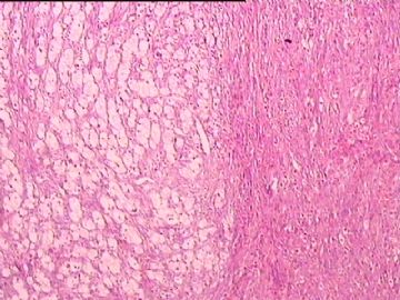
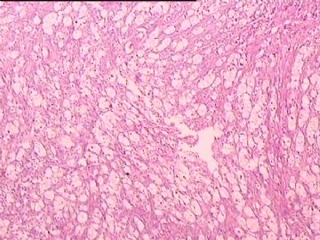
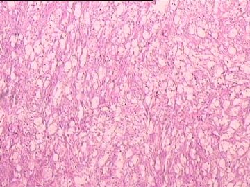
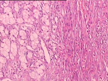
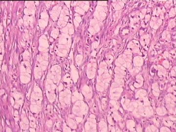
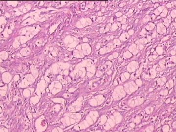
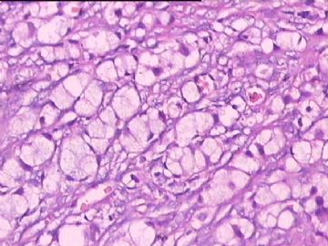
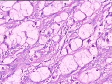
Before jumping on diagnosis, I would like to ask more information from the 楼主。
1) Gross appearance: Is there a mass? Is the color of the mass yellow and soft or like you described "grey-white and firm" ? Is there any cystic areas in this ovary? Is lesion unilateral or bilateral?
2) Microscopically, is this all you see or any other areas with different structure and hisopathology?
3) Most importantly, what is clinical manifestation? Does patient have any androgenic or estrogenic signs? Any cushing's signs? What is prompt for this oophrectomy? Any stomach symptoms?
Knowing these information will be helpful to make your differential diagnoses. Now based solely on morphologic alone, you may include or add the following to your diagnosis:
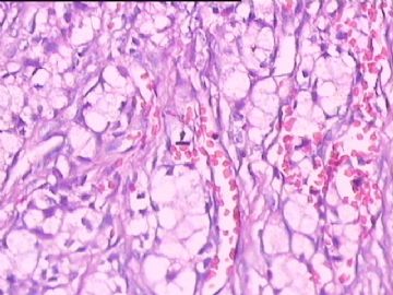
1) Steroid cell tumor, NOS: I like to know if patient has any androgenic or estrogenic or Cushing's presentation since more than 50% patients of this tumor will have such clinical manifestation. IHC should be positive both for inhibin and vimentin. The gross appearance should be YELLOW in this case.
2) Thecoma or fibrothecoma: I like to know if there is solid fiboma-like areas. Sometimes rare adult-type granulosa cell tumor can simulate thecoma-like change and should be aware of it. In that case, Retic staining is helpful in differentiating these two entities.
3) Krukenburg tumor: this tumor can be presented in variety of morphology including sth like this case with clear cell changes. Sometimes signet ring cells are very cryptic and deceptive. Usually Krukenberg tumor is bilateral, but it can be unilateral too, especially on right side. A mucin staining and AE1/3 should be applied to rule out this cancer.
4) Other rarely seen tumor, such as Leydig cell tumor, metastatic renal cell carcinoma, metaslatic melanoma and so on should be also in your DDx.
5) Occasionally a ovarian luteoma with focal hemorrhage and evolving histiocytes aggregation can mislead us to a true neoplastic diagnosis. But luteoma is usually related to recent pregnancy and patient's history should give clue of it.
So the bottom line for this case is that we need more information to fuel the expanded discussion. As I emphasized in the past, it is very very important for us all here to establish a solid and logic thought process which leads us to the right diagnosis, instead of just "bet" on a diagnosis based on limited photos and fields. I hope this is helpful.
abin译:
在试图诊断之前,我想请楼主再提供一些信息。
1)大体:有肿块吗?肿块是黄色、软还是你描述的“灰白、硬”?卵巢有囊性区吗?病变是单侧还是双侧?
2)镜下:这是你的全部所见,或者其它区域没有不同结构和组织学表现?
3)最重要的是,临床表现?患者有高雄激素或高雌激素表现吗?有Cushing综合征表现吗?为什么切除卵巢?有胃症状吗?
知道这些信息将有助于鉴别诊断。现在仅根据形态学表现分析如下,你可以包括或增加到你的诊断之中:
1) Steroid细胞瘤,NOS: 我想知道患者是否有高雄激素或高雌激素或Cushing综合征表现,因为这种肿瘤超过50%患者会有临床表现。IHC检测inhibin 和vimentin均为阳性。大体表现应该是黄色。
2) 卵泡膜瘤或纤维卵泡膜瘤: 我想知道是否有实性的纤维瘤样区域。有时罕见的成人型粒层细胞瘤或能假冒卵泡膜瘤样改变,应该小心。网染有助于区分二者。
3) Krukenburg瘤: 可表现不同形态,包括本例这样的透明细胞改变。有时印戒细胞非常隐蔽、具有欺骗性。通常Krukenburg瘤是双侧,但也可单侧,特别是右侧。粘液染色和AE1/3可用于排除它。
4) 其它罕见肿瘤,如Leydig细胞瘤、转移性恶黑等也应鉴别。
5) 偶尔,卵巢黄体瘤伴局灶出血和继发组织细胞聚集可能误导我们作出真性肿瘤的诊断。但是黄体瘤通常与最近妊娠有关,患者病史会提供线索。
因此本例的底线是,我们需要更多信息,以助扩大讨论。正如我以前强调的,我们最重要的是确立坚实的、逻辑性的诊断思路,从而引导我们作出正确诊断,而不是仅仅根据有限的图像和视野“赌”一个诊断。希望这样有所帮助。

- 不坠青云之志,长怀赤子之心
在作出诊断之前,我想从楼主处获得更多的信息
1)大体表现:此肿瘤是实性的吗?色黄,质软还是如楼主所述的灰白质实?有囊性区域吗?单侧发生还是双侧?
2)镜下表现:完全是如你所提供的图一样的表现吗?有没有其他不同的结构和镜下表现?
3)最重要的是,患者有何临床表现?有没有男性化或者高性激素的表现?有没有Cushing's综合症的表现?此种表现是瞬发的还是逐渐改变的?有没有胃疾病的表现?
以上这些信息有助于我们做出正确的鉴别诊断。仅从你所提供的图片来分析,可以从以下方面去考虑诊断:
1)类固醇细胞瘤,非特异性:我想知道患者有没有男性化特征或高性激素表现或者Cushing's综合症表现?大约有超过50%的此种肿瘤患者会有此临床表现。IHC显示inhibin和vim阳性。大体表现应该是黄色。
2)卵泡膜细胞瘤或卵泡膜纤维瘤:在此肿瘤中是否有实性的纤维化区域?必须注意,有时极少数adult-type粒层细胞瘤可以发生刺激发生卵泡膜样改变,在这样的病例中,网址纤维染色有助于鉴别此两种肿瘤。
3)Krukenburg瘤:此肿瘤形态学具有多样性,比如在此例中的类似透明细胞样改变的区域。有时候印戒细胞形态学不典型,具有一定的迷惑性。通常Krukenburg瘤双侧发生,但是也有单侧发生的病例,特别是右侧。粘蛋白染色和免疫组化的AE1/3有助于鉴别此类肿瘤。
4)其他少见肿瘤,比如间质细胞瘤,转移性肾细胞癌,转移性黑色素瘤等。
5)有时候,卵巢黄体瘤伴有局灶性出血,大量组织细胞聚集时候会误导我们诊断为新生物。但是黄体瘤患者通常最近有妊娠史,这一点会给提供给我们一定的线索。
最后还是要强调一点,对于此例我们还需要更多的信息来进一步供我们讨论。正如上面我所强调的,我们大家
都需要有一个坚实的逻辑思考过程来指导我们得出正确的诊断,而不是仅仅在所提供有限的图像和有限的镜下
所见基础上表示支持某种诊断。希望对大家有所帮助……

- 赚点散碎银子养家,乐呵呵的穿衣吃饭
| 以下是引用杨斌在2009-4-24 22:58:00的发言:
Before jumping on diagnosis, I would like to ask more information from the 楼主。 1) Gross appearance: Is there a mass? Is the color of the mass yellow and soft or like you described "grey-white and firm" ? Is there any cystic areas in this ovary? Is lesion unilateral or bilateral? 2) Microscopically, is this all you see or any other areas with different structure and hisopathology? 3) Most importantly, what is clinical manifestation? Does patient have any androgenic or estrogenic signs? Any cushing's signs? What is prompt for this oophrectomy? Any stomach symptoms? Knowing these information will be helpful to make your differential diagnoses. Now based solely on morphologic alone, you may include or add the following to your diagnosis: 1) Steroid cell tumor, NOS: I like to know if patient has any androgenic or estrogenic or Cushing's presentation since more than 50% patients of this tumor will have such clinical manifestation. IHC should be positive both for inhibin and vimentin. The gross appearance should be YELLOW in this case. 2) Thecoma or fibrothecoma: I like to know if there is solid fiboma-like areas. Sometimes rare adult-type granulosa cell tumor can simulate thecoma-like change and should be aware of it. In that case, Retic staining is helpful in differentiating these two entities. 3) Krukenburg tumor: this tumor can be presented in variety of morphology including sth like this case with clear cell changes. Sometimes signet ring cells are very cryptic and deceptive. Usually Krukenberg tumor is bilateral, but it can be unilateral too, especially on right side. A mucin staining and AE1/3 should be applied to rule out this cancer. 4) Other rarely seen tumor, such as Leydig cell tumor, metastatic renal cell carcinoma, metaslatic melanoma and so on should be also in your DDx. 5) Occasionally a ovarian luteoma with focal hemorrhage and evolving histiocytes aggregation can mislead us to a true neoplastic diagnosis. But luteoma is usually related to recent pregnancy and patient's history should give clue of it. So the bottom line for this case is that we need more information to fuel the expanded discussion. As I emphasized in the past, it is very very important for us all here to establish a solid and logic thought process which leads us to the right diagnosis, instead of just "bet" on a diagnosis based on limited photos and fields. I hope this is helpful. abin译: 在试图诊断之前,我想请楼主再提供一些信息。 2)镜下:这是你的全部所见,或者其它区域没有不同结构和组织学表现? 3)最重要的是,临床表现?患者有高雄激素或高雌激素表现吗?有Cushing综合征表现吗?为什么切除卵巢?有胃症状吗? 知道这些信息将有助于鉴别诊断。现在仅根据形态学表现分析如下,你可以包括或增加到你的诊断之中: 1) Steroid细胞瘤,NOS: 我想知道患者是否有高雄激素或高雌激素或Cushing综合征表现,因为这种肿瘤超过50%患者会有临床表现。IHC检测inhibin 和vimentin均为阳性。大体表现应该是黄色。 2) 卵泡膜瘤或纤维卵泡膜瘤: 我想知道是否有实性的纤维瘤样区域。有时罕见的成人型粒层细胞瘤或能假冒卵泡膜瘤样改变,应该小心。网染有助于区分二者。 3) Krukenburg瘤: 可表现不同形态,包括本例这样的透明细胞改变。有时印戒细胞非常隐蔽、具有欺骗性。通常Krukenburg瘤是双侧,但也可单侧,特别是右侧。粘液染色和AE1/3可用于排除它。 4) 其它罕见肿瘤,如Leydig细胞瘤、转移性恶黑等也应鉴别。 5) 偶尔,卵巢黄体瘤伴局灶出血和继发组织细胞聚集可能误导我们作出真性肿瘤的诊断。但是黄体瘤通常与最近妊娠有关,患者病史会提供线索。 因此本例的底线是,我们需要更多信息,以助扩大讨论。正如我以前强调的,我们最重要的是确立坚实的、逻辑性的诊断思路,从而引导我们作出正确诊断,而不是仅仅根据有限的图像和视野“赌”一个诊断。希望这样有所帮助。 |
-
I agree with Dr. yang's excellent analysis. Pathologists should also know as much information as you can, then think over the possible different diangosis, even though for these easy cases. For the difficult cases you need to dicide which stains can help you to make the diagnosis ( or rule out). You need to think your cases again when you have your IHC. You can release your cases if you are sure. Otherwise you may need more IHC or consult other pathologists.

