| 图片: | |
|---|---|
| 名称: | |
| 描述: | |
- B1781breast lesion
| 姓 名: | ××× | 性别: | female | 年龄: | 54 |
| 标本名称: | Breast lesion | ||||
| 简要病史: | |||||
| 肉眼检查: | |||||
Good morning, my colleagues:
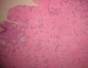
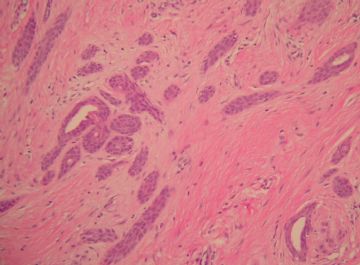
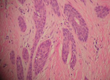
This is an interesting case of breast lesion. I would like to share with you for educational purpose. My fellow and I will publish a short review of this entity in the future issue of Archives of Pathology and Laboratory Medicine.
This is a 54-year-old female with history of breast nodule measuring 2.5 x 2 x 1.8 cm. Skin over the nodule shows crusting and ulceration. Mammography demonstrates a mass in the subareolar region with dense and irregular outline, spicular formation and foci of microcalcifications. Ultrasonography shows an ill-defined mass with heterogeneous internal echoes. Radiographic findings suggest malignancy.
-
本帖最后由 于 2009-04-20 20:36:00 编辑
-
本帖最后由 于 2009-05-15 23:58:00 编辑
Good morning, my colleagues:
This is an interesting case of breast lesion. I would like to share with you for educational purpose. My fellow and I will publish a short review of this entity in the future issue of Archives of Pathology and Laboratory Medicine.
This is a 54-year-old female with history of breast nodule measuring 2.5 x 2 x 1.8 cm. Skin over the nodule shows crusting and ulceration. Mammography demonstrates a mass in the subareolar region with dense and irregular outline, spicular formation and foci of microcalcifications. Ultrasonography shows an ill-defined mass with heterogeneous internal echoes. Radiographic findings suggest malignancy.
名位同行,早上好:
这是一例有趣的乳腺病例,做为教学目的我想与你们分享这一病例。我与我的进修生将在下一期Archives of Pathology and Laboratory Medicine杂志上发表一篇这种病变的短的回顾性研究。
这是一个54岁的女性,乳腺有一2.5*2*1.8cm肿块。肿块表面皮肤溃疡、结痂。乳房影像学显示在乳晕下区域有一边界不规则的高密度肿块,毛刺状外形,灶性微钙化。超声检查显示一内部回声不均质的界限不清楚的肿块。放射学结果考虑恶性肿瘤。
搬个马扎学习......

- 我思故我在! know something about everything,know everything about something.
-
liguoxia71 离线
- 帖子:4174
- 粉蓝豆:3122
- 经验:4677
- 注册时间:2007-04-01
- 加关注 | 发消息


