| 图片: | |
|---|---|
| 名称: | |
| 描述: | |
- 求助:女,46岁,右耳后腮腺区约2.5cm肿块。
-
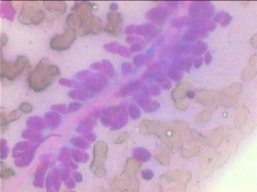
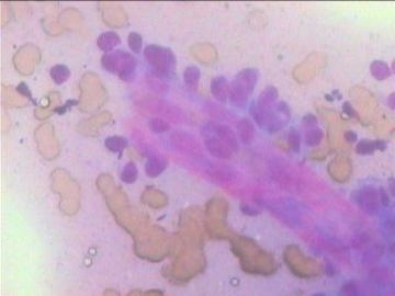
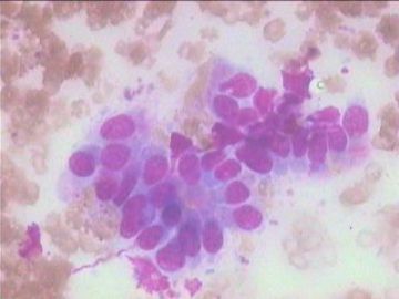
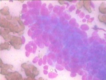
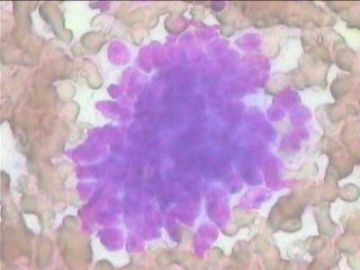
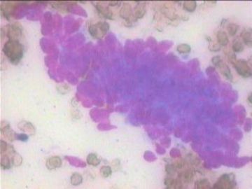
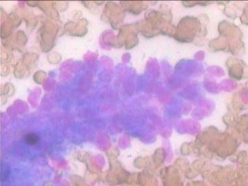
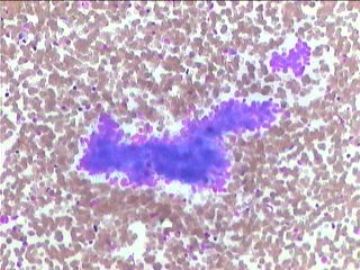
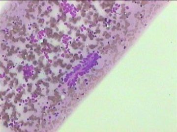
非常感谢liguoxia71和陈博士回帖,由于我的显微镜有些差,图象捕捉系统是盗版的水货,所以发上来的图片有些模糊不清,在此深表歉意!本例当时穿刺的时候,病人有意隐瞒了病史和治疗史,只是口头告诉有鼻咽癌史,我一再追问,她都没有进一步深说,当时她情绪很差,所以我就没有再追问。穿刺部位是右腮腺区,镜下背景中未找到淋巴细胞,见部分感觉上皮来源的细胞团,极象鼻咽等来源的非角化鳞癌细胞(见图5、6),但又可见如图1、2、7、8、9等感觉不象上述非角化型鳞癌细胞样细胞团,且排列也不大象,也可见如图3样柱状腺皮腺腔样排列的细胞团,特别是镜下未见淋巴细胞。所以我当时倾向的诊断是:1、转移性非角化性鳞癌可能性大;2、腮腺原发性肿瘤(上皮-肌上皮癌不能排除)不能排除;建议手术活检。感觉本例有点意思,于是昨天又给患者家属打电话了,建议无论如何要来医院手术活检或切除,她们告之曾在某医院鼻咽镜活检诊断鼻咽癌且行过多次放疗,眼下是否手术,只有家属间商议后再做决定;而且,要她们去那家医院借切片,估计非常困难,因为涉及到某些敏感事情;所以,陈博士建议比较本次与以前切片细胞学表现,估计很难办到,眼下唯一可以施行的是动员其手术。因此,我将试着再建议让她来院手术及其它相关检查,在此再次希望各位网友给些建议,谢谢!
Very interesting case. Unfortunately, the quality of the slide is suboptimal. If all the slides look like this, it is definitely a neoplasm. You need to get the history of what exactly is her previous malignancy, if possible, compare the morphology of that. It is very important to differentiate primary salivary gland neoplasm vs. metastasis, since the treatment will be very different.
My approach to salivary gland FNA is that if a tumor is not neatly fit into any known subtype of salivary gland neoplasm, don't force yourself into one specific diagnosis, if it is primary, ask the surgon to take it out. Salivary gland FNA can be very difficult and have a lot of overlapping features. Cytologists are usually good at differentiating neoplasm vs. non-neoplastic process and low-grade vs. high-grade maliganant neoplasm, but have problems with a specific diagnosis.
Back to this case, I not only see some papillary structure but also see some glandular or tubular formation, I don't see a distinct myoepithelial component. I favor this is an epithelial neoplasm, maybe just adenocarcinoma.

