| 图片: | |
|---|---|
| 名称: | |
| 描述: | |
- 50岁,绝经
-
本帖最后由 于 2009-04-05 01:28:00 编辑
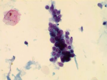
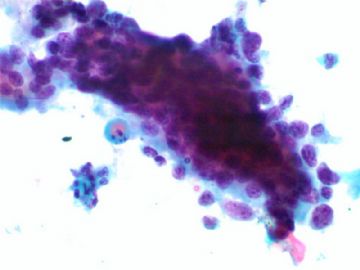
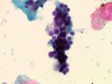
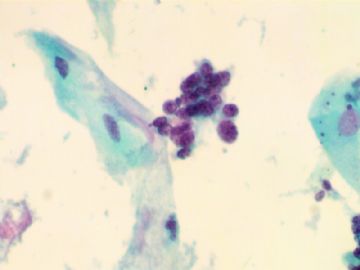
Very interesting case. I showed the photos to Dr. Austin (he is our director of cytopathology and previous president of American Society of Cytopathology) and other cytopathologist. All of our three have similar oppinion.
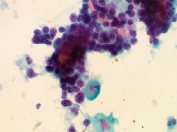
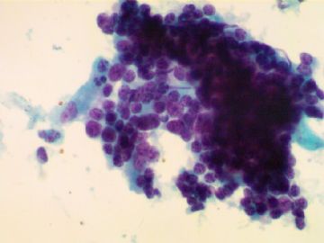
1. We cannot report malignant,adenocarcinoma, or HSIL, based on the cytology above.
2. These cells look like endometrial cell orgin
3. Agree with Dr. Yu these cells mostly are endometrial cells from low uterin segment due to the sampling. There is mild cytologic atypia in high power. Few cells are large than others (two times)---pleomorphism.
The question is what we should call, endometrial cells in 绝经 (even though mostly the presence of the cells were due to the sampling) or atypical glandular cells.
The bottomline is that the women should have tissue sampling. Based on ASCCP guidline the women should have tissue sampling even we report normal endometrial cells in 绝经 women.
I may call normal endometrial cells if I take the test.
We all three agree to call atypical glandular cells for this true clinical practice. I hope to make sure the women get tissue sampling, even though the result turns out to be negative in histologic specimen. Remember that 70-80% of women with AGC Pap will have no any endometrial or endocervical lesions in histologic follow-up. Also the diagnosis of endometrioid adenocarcinoma in histology is often based on the architecture, but not cytology.
I did a lot AGC study and reviewed many AGC slides and have three publications in AGC. The more I study, the more I am confuded.
Just for your reference.
-
jiangxiaoyu 离线
- 帖子:978
- 粉蓝豆:15
- 经验:1226
- 注册时间:2007-10-31
- 加关注 | 发消息
-
Direct sampling of endometrium. Don't have to report. Geographic irregular groups of small piled up cells with very scant cytoplasm. Spontaneously exfoliated endometrial groups are smaller size with round contour, have to report in postmaneupausal women. Don't mistake for HSIL. What is AIG? American International Group in Wall Street who handed out fat bonuses to high executives using tax payers' sweat money?

