共3页/50条首页上一页123下一页尾页
回复:50 阅读:8713
B1770Breast fibroepithelial lesions (cqz 15)

cqzhao 离线

帖子:5518
粉蓝豆:1071
经验:5755
注册时间:2008-09-29
加关注  |  发消息
楼主 发表于 2009-03-07 12:55|举报|关注(0)
浏览排序[ 顺序 逆序 楼主 支持 精彩 ]  快捷回复
姓    名: ××× 性别:   年龄:  
标本名称:  
简要病史:  
肉眼检查:  

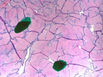
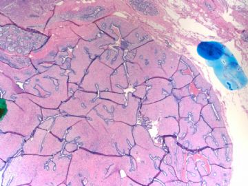
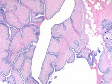
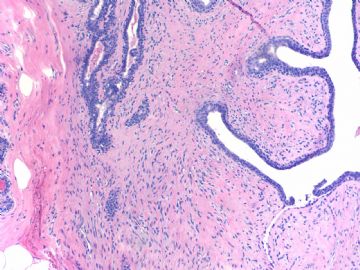
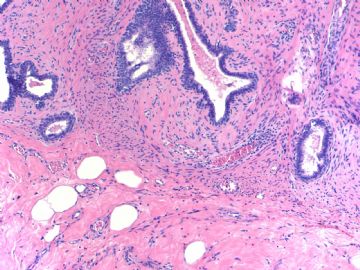
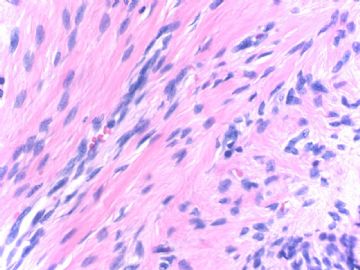
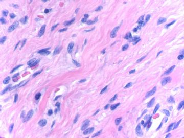
I notoced there are some cases of fibroepithelial lesions (biphasic tumor)in our website. I send this case to open the discussion.

Fibroepithelial tumors including fibroadenoma (FA) and phyllodes tumors (PT). The definition and classification of PT are not very agreeable among pathologists. However, they are most common classified as bengin, borderline, or malignant suggested by WHO. Some pathologists use terms of benign, low grade maligant and high grade malignant. These two systems are very similar, borderlin equal to low grade malignant.

Hope you can give your diagnosis and mention why. I assume people may have different oppinions. It is ok.  We just discuss the case.

  • Breast fibroepithelial lesions (cqz 15)图1
    图1
  • Breast fibroepithelial lesions (cqz 15)图2
    图2
  • Breast fibroepithelial lesions (cqz 15)图3
    图3
  • Breast fibroepithelial lesions (cqz 15)图4
    图4
  • Breast fibroepithelial lesions (cqz 15)图5
    图5
  • Breast fibroepithelial lesions (cqz 15)图6
    图6
  • Breast fibroepithelial lesions (cqz 15)图7
    图7
标签:乳腺纤维腺瘤 叶状肿瘤
0
添加参考诊断
×参考诊断
  

有福不在忙 离线

帖子:2161
粉蓝豆:42
经验:3448
注册时间:2008-11-22
加关注  |  发消息
41 楼    发表于2009-03-15 17:28:00举报|引用
返回顶部 | 快捷回复
 第一例我同意交界性PTs;第二例我认为是FA或良性PTs。
0
回复
signature
有福不在忙

abin 离线

帖子:7416
粉蓝豆:6014
经验:11787
注册时间:2006-10-08
加关注  |  发消息
42 楼    发表于2009-03-17 01:00:00举报|引用
返回顶部 | 快捷回复

 第1例,同第4楼。

第2例:如果图1和6反映病变的边界情况,它的边界是不清楚的,没有包膜,不好直接诊断纤维腺瘤。

周围病变似乎有硬化性腺病和小叶增生。因此总体上考虑“乳腺增生症伴纤维腺瘤形成趋势”。因为它无指状突起,无亲上皮性生长,无核分裂,无明显异型性,间质密度也不高,所以不考虑叶状肿瘤。

0
回复
signature

华夏病理/粉蓝医疗

为基层医院病理科提供全面解决方案,

努力让人人享有便捷准确可靠的病理诊断服务。


cqzhao 离线

帖子:5518
粉蓝豆:1071
经验:5755
注册时间:2008-09-29
加关注  |  发消息
43 楼    发表于2009-04-10 10:22:00举报|引用
返回顶部 | 快捷回复
 About case 2: Patient had history of phyllodes tumor several years ago (outside hospital).  So this is local recurrence of benign phyllodes. It is difficult to predict the prognosis for phyllodes. The major concern is local recurrence. Distant metastases are rare.Complete excision with wide free margin is the main treatment. One large review study indicated the chance of local recurrence was 21%, 46%, and 65% for the patients with benign, borderline, and malignant phyllodes.
0
回复

cqzhao 离线

帖子:5518
粉蓝豆:1071
经验:5755
注册时间:2008-09-29
加关注  |  发消息
44 楼    发表于2009-04-10 10:28:00举报|引用
返回顶部 | 快捷回复
 Elizabeth : glad to see you at Hong Kong. Could you summarize the distinguishing features for benign, borderline and malignant phyllodes tumors? Thanks, cz
0
回复

小荷 离线

帖子:8546
粉蓝豆:97
经验:12725
注册时间:2006-09-11
加关注  |  发消息
45 楼    发表于2009-04-10 22:13:00举报|引用
返回顶部 | 快捷回复
 赵老师您见到  Elizabeth了啊。能否发张照片上来啊,我们也想见见啊
0
回复
signature
没有完美的个人,只有完美的团队

cqzhao 离线

帖子:5518
粉蓝豆:1071
经验:5755
注册时间:2008-09-29
加关注  |  发消息
46 楼    发表于2009-04-11 01:09:00举报|引用
返回顶部 | 快捷回复
 I do not have. you can ask Elizabeth send you a photo. A nice @ beautiful Chinese lady just like you.
0
回复

qianxun 离线

帖子:45
粉蓝豆:2
经验:54
注册时间:2009-04-10
加关注  |  发消息
47 楼    发表于2009-04-11 05:24:00举报|引用
返回顶部 | 快捷回复

 For case #1 (09-3-7)

  Phyllodes tumor, histologically benign.

Comment:  small encapsulated lesion, low stromal cellularity , No mitosis (less than 0-4 mitotic figures/10 HPF) seen on the pictures althought scattered stromal cells showed mild muclear atypia.   

0
回复

天山望月 离线

帖子:4902
粉蓝豆:83
经验:5092
注册时间:2007-02-03
加关注  |  发消息
48 楼    发表于2009-03-21 16:03:00举报|引用
返回顶部 | 快捷回复
本帖最后由 于 2009-03-21 18:05:00 编辑

FA和PT的鉴别,不足之处,请专家指导,谢谢!

   FA  PT
发病年龄

 多在20-30岁

 多在45岁以上

肿瘤性状

边界

包膜

切面

质地


 清

灰白色,有光泽,编织状


 稍清

灰瓷白色,有光泽,细腻

脆,穿刺组织常较碎。


 镜下  上皮和间质混合性增生,常比例相当,或上皮成分多于间质,每10倍视野均可见腺上皮。间质可有粘液样变,细胞稀疏,无异型性,极少核分裂。  上皮和间质混合性增生,间质增生明显,每10倍视野可见无腺上皮成分区。间质可有粘液样变,细胞密集程度大于FA,有异型性,可见核分裂。
 预后  手术切除,很少复发,预后好。  良性有一定的复发率,恶性可有复发和转移。


0
回复
signature
广州金域病理

天山望月 离线

帖子:4902
粉蓝豆:83
经验:5092
注册时间:2007-02-03
加关注  |  发消息
49 楼    发表于2009-03-21 18:18:00举报|引用
返回顶部 | 快捷回复

 仔细观察 Case2:图1中,从图中看,病变无包膜,部分为纤维腺病样(右上),(左下)部分粘液样背景中增生的导管,结节状,但无明显分叶状结构。20倍视野可见多个腺体,间质细胞稀疏,无异型性,未见核分裂。

综合考虑:此例为纤维腺病伴局部纤维腺瘤样增生。

0
回复
signature
广州金域病理

alading1999 离线

帖子:63
粉蓝豆:31
经验:562
注册时间:2009-01-17
加关注  |  发消息
50 楼    发表于2009-03-21 22:53:00举报|引用
返回顶部 | 快捷回复
 第一例 良性叶状肿瘤, 第二例 幼年型纤维腺瘤?
0
回复
回复:50 阅读:8713
共3页/50条首页上一页123下一页尾页
【免责声明】讨论内容仅作学术交流之用,不作为诊疗依据,由此而引起的法律问题作者及本站不承担任何责任。
快速回复
进入高级回复
您最多可输入10000个汉字,按 "Ctrl" + "Enter" 直接发送
搜索回复/乘电梯 ×
按内容
按会员
乘电梯
合作伙伴
友情链接