| 图片: | |
|---|---|
| 名称: | |
| 描述: | |
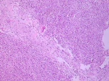
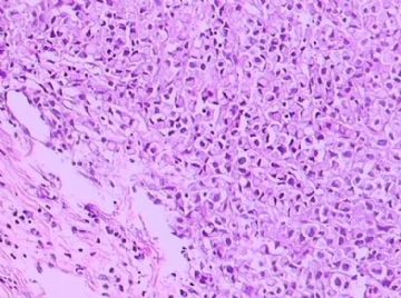
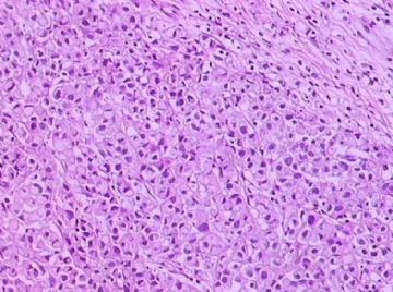
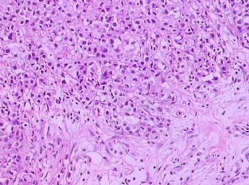
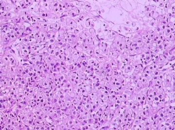
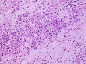
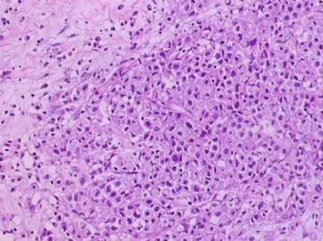
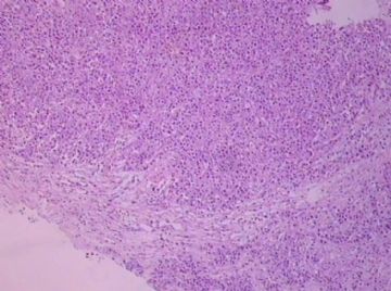
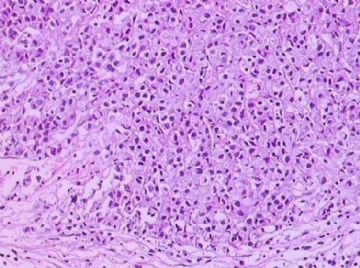
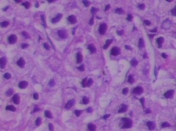
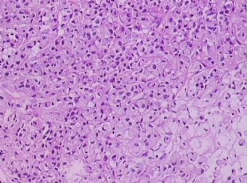
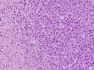
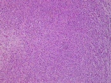
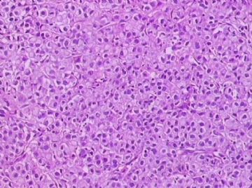
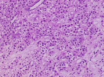
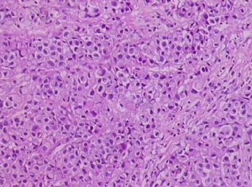
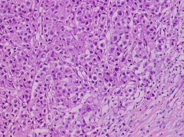
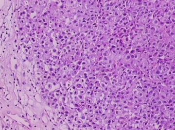
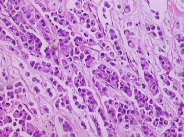
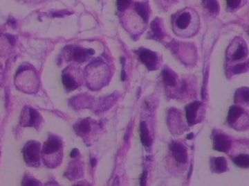
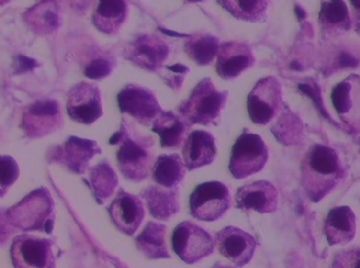
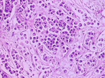
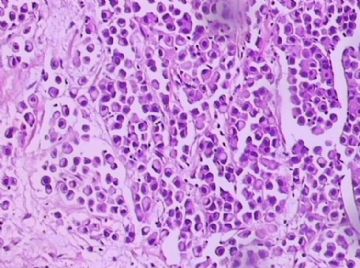
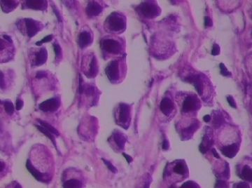
- B1762乳腺印戒细胞癌?
-
shn-821128 离线
- 帖子:277
- 粉蓝豆:3
- 经验:277
- 注册时间:2008-11-02
- 加关注 | 发消息
关于印戒细胞癌,WHO2003只有一段。最后一句还是翻译错的。
Signet ring cell carcinoma (8490/3)
Signet ring cell carcinomas are of two types. One type is related to lobular carcinoma and is characterized by large intracytoplasmic lumina which compress the nuclei towards one pole of the cell. Their invasive component has the targtoid pattern of classical lobular carcinoma. The other type is similar to diffuse gastric carcinoma, and is characterized by acidic mucosubstances that fill the cytoplasm and dislodge the nucleus to one pole of the cell. This type of signet ring cell carcinoma can be seen in association with the signet ring cell variant of DCIS.
印戒细胞癌有2型。其一与小叶癌有关,特征为瘤细胞浆内含大空腔并将核挤到边缘。浸润成分具有经典小叶癌的靶环状结构。另一型与弥漫性胃癌相似,特征是胞浆内含嗜酸性黏液物质并将核挤到边缘。后者可能与DCIS的印戒细胞亚型相伴随。

华夏病理/粉蓝医疗
为基层医院病理科提供全面解决方案,
努力让人人享有便捷准确可靠的病理诊断服务。
-
本帖最后由 于 2009-03-08 21:07:00 编辑
Seem every one agree this is a signet ring cell ca, breast primary (one type of lobular ca) or metastatic ca. Generally it is a primary tumor. But we can do IHC, CK7, ck20, ER, CDX2 pr to rule out GI primary.
Often
Breast: ck7+/ck20-, ER+, CDx2-
GI: CK7-/ck20+, er-, CDx2+
In fact GI: gastric + colonic origin
E-cadherin seems not very useful (see below article). No lot study for p120.
abin译:似乎所有人都同意印戒细胞癌,乳腺原发(小叶癌的一个亚型)或转移癌。它一般是原发肿瘤。可以做CK7, ck20, ER, CDX2 pr 排除消化道转移(胃和结肠)。
乳腺:ck7+/ck20-, ER+, CDX2-
消化道:CK7-/ck20+, ER-, CDX2+
E-cadherin似乎没有太大帮助(见以下论文)。p120研究不多。
-
Am J Clin Pathol. 2004 Jun;121(6):884-92.
-
Immunohistochemical characterization of signet-ring cell carcinomas of the stomach, breast, and colon.
Division of Pathology, City of Hope National Medical Center, Duarte, CA 91010, USA.
We studied the immunophenotype of signet-ring cell carcinoma (SRCC) of the stomach (30 cases), breast (21 cases), and colon (9 cases) with the following expression patterns: (1) breast: consistent, MUC1 (21 [100%]), cytokeratin (CK) 7 (20 [95%]), estrogen receptor (ER; 17 [81%]); infrequent, E-cadherin (6 [29%]), MUC2, MUC5AC, CK20 (1 [5%] each); negative, CDX2 and hepatocyte paraffin 1 (Hep Par 1; 0 [0%] each); (2) gastric: frequent, CDX2 (27 [90%]) and Hep Par 1 (25 [83%]); variable, E-cadherin and CK20 (17 [57%] each), MUC2 and MUC5AC (15 [50%] each), MUC1 (5 [17%]); negative, ER (0 [0%]); and (3) colon: frequent, MUC2 (9 [100%]), CDX2 and MUC5AC (8 [89%] each); infrequent or negative, MUC1 (3 [33%]), Hep Par 1 (2 [22%]), ER (0 [0%]). Immunohistochemical staining distinguished breast from gastric SRCC (ER, MUC1, Hep Par 1, CDX2) and colon SRCC (ER, CDX2, MUC2, and MUC5AC). Gastric and colon SRCCs showed a similar staining pattern for antibodies tested except for Hep Par 1 and CDX2 (gastric, 83% Hep Par 1 positivity and heterogeneous, weak, patchy CDX2 nuclear staining; colon, 22% Hep Par 1 positivity and homogeneous, strong, diffuse CDX2 nuclear staining). About half of the cases of gastric SRCC expressed MUC2 and MUC5AC, whereas virtually all cases of colon SRCC expressed them.
-
Am J Surg Pathol. 2008 Aug;32(8):1182-9.
-
Immunohistochemical staining of Reg IV and claudin-18 is useful in the diagnosis of gastrointestinal signet ring cell carcinoma.
Department of Molecular Pathology, Hiroshima University Graduate School of Biomedical Sciences, Hiroshima, Japan.
Signet-ring cell carcinoma (SRCC) is a unique subtype of adenocarcinoma that is characterized by abundant intracellular mucin accumulation and a crescent-shaped nucleus displaced toward one end of the cell. Identification of an SRCC's primary site is important for better planning of patient management because the treatment and prognosis differs markedly depending on the origin of the SRCC. In the present study, we analyzed the immunohistochemical characteristics of 94 cases of SRCC, including 21 cases of gastric SRCC, 16 of colorectal SRCC, 10 of breast SRCC, and 47 of pulmonary SRCC, with antibodies against Reg IV and claudin-18, which we previously identified as gastric cancer-related genes. We also tested known markers cytokeratin 7, cytokeratin 20, MUC2, MUC5AC, caudal-related homeobox gene 2 (CDX2), thyroid transcription factor-1, mammaglobin, gross cystic disease fluid protein 15, and estrogen receptor. All 21 cases of gastric SRCC and 16 cases of colorectal SRCC were positive for Reg IV, and the remaining SRCCs were negative. Eighteen of 21 (86%) gastric SRCCs and 6 of 16 (38%) colorectal SRCCs were positive for claudin-18, whereas another SRCCs were negative. In conclusion, Reg IV staining and claudin-18 staining can aid in diagnosis of gastrointestinal SRCC.
-
Int J Gynecol Pathol. 2004 Jan;23(1):45-51.
-
Signet-ring stromal tumor of the ovary: clinicopathologic analysis and comparison with Krukenberg tumor.
Department of Gynecologic and Breast Pathology, Armed Forces Institute of Pathology, Washington, DC 20306-6000, USA. rvang1@yahoo.com
Signet-ring stromal tumor is a rare ovarian neoplasm that can mimic Krukenberg tumor because of the presence of signet-ring cells in both tumors. The clinicopathologic features of three signet-ring stromal tumors, one of which has been previously reported, were analyzed and compared with 10 Krukenberg tumors. Patients with signet-ring stromal tumor ranged in age from 34 to 41 years (mean: 36.7 years). All signet-ring stromal tumors were unilateral and stage IA, whereas 60% and 40% of Krukenberg tumors were bilateral or associated with extraovarian tumor, respectively. The signet-ring stromal tumors were devoid of epithelial differentiation (glands, nests, cords), whereas all of the Krukenberg tumors contained these epithelial structures at least focally. In contrast to signet-ring stromal tumors, the signet-ring cells of Krukenberg tumors were positive for periodic acid-Schiff with diastase and cytokeratins but negative for vimentin. The patients with signet-ring stromal tumors were alive without disease at follow-up interval of 1 month to 17.4 years (mean: 7.4 years). In summary, signet-ring stromal tumor is a rare, benign, ovarian tumor that may be mistaken for Krukenberg tumor. Although the combination of operative and histopathologic findings allow their distinction, histochemical and immunohistochemical stains may also be useful.
-
APMIS. 2000 Jun;108(6):467-72.
-
The role of cytokeratins 20 and 7 and estrogen receptor analysis in separation of metastatic lobular carcinoma of the breast and metastatic signet ring cell carcinoma of the gastrointestinal tract.
Department of Pathology and Clinical Cytology, Central Hospital, Falun, Sweden. tibor.tot@ltdalarna.se
Metastatic signet ring cell carcinomas of unknown primary site can represent a clinical problem. Gastrointestinal signet ring cell carcinomas and invasive lobular carcinomas of the breast are the most common sources of these metastases. Immunohistochemical algorithms have been successfully used in the search for the unknown primary adenocarcinomas. In the present study a series of primary invasive lobular breast carcinomas (79 cases) and their metastases and a series of gastrointestinal signet ring cell carcinomas (22 primary and 13 metastases) were stained with monoclonal antibodies for cytokeratin (CK) 20 and CK7 and for estrogen receptors (ER). The staining was evaluated as negative (no staining), focally (less than 10% of the tumor cells stained) or diffusely positive. All the primary and metastatic gastrointestinal signet ring cell carcinomas proved to be CK20 positive, while only 2/79 (3%) of the primary and 1/21 metastatic lobular carcinomas (5%) stained positively for this CK. None of the gastrointestinal carcinomas and the majority of the lobular carcinomas expressed ER. The majority of the tumors were CK7+. Using CK20 alone, 33 of 34 metastases could be properly classified as gastrointestinal (CK20+) or mammary (CK20-). ER identified 31/34 of breast cancer metastases. By combining the results of CK20 and ER staining all the metastases could be properly classified as the CK20+/ER- pattern identified all the gastrointestinal tumors.
-
Mod Pathol. 1993 Sep;6(5):516-20.
-
Signet ring variant of lobular carcinoma of the breast: a clinicopathologic and immunohistochemical study.
Department of Pathology, Henry Ford Hospital, Detroit, Michigan.
Signet ring carcinoma of the breast often metastasizes to gastrointestinal tract and female genital tract. We report clinicopathologic features of 10 breast carcinomas with signet ring features, five of which had unusual metastatic patterns. The primary breast tumor in all these cases was lobular carcinoma. Although signet ring cells were prominent in metastatic sites, the primary tumor lacked signet ring cells in two cases. A linitis plastica-like presentation and presence of signet ring cells in gastric metastases raised a strong possibility of primary gastric carcinoma in three cases. The monoclonal antibody to gross cystic disease fluid protein (GCDFP-15) was positive in signet ring cell-rich areas in the primary breast tumor (8/10) and/or in the metastases in all cases. For comparison we studied GCDFP-15 immunoreactivity in 10 infiltrating lobular and 10 infiltrating ductal breast carcinomas with no obvious signet ring cells, and in 14 signet ring carcinomas from other sites (10 gastric, 2 prostatic, 2 colonic). The gastric, colonic, and one prostatic signet ring carcinoma were nonreactive. One prostatic signet ring carcinoma exhibited focal but unequivocal positivity with GCDFP-15. The cases of this report reinforce the concept that signet ring carcinoma of the breast is usually a variant of lobular carcinoma and not a distinct entity. Signet ring cell predominance in metastases, even in the absence of signet ring cells in the primary tumor, attest to the morpho-functional heterogeneity of lobular carcinoma. GCDFP-15 is a sensitive marker for signet ring breast carcinoma and a very useful adjunct tool in the diagnosis of metastatic signet ring carcinoma of mammary origin

