| 图片: | |
|---|---|
| 名称: | |
| 描述: | |
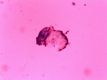
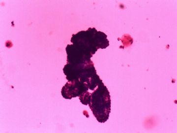
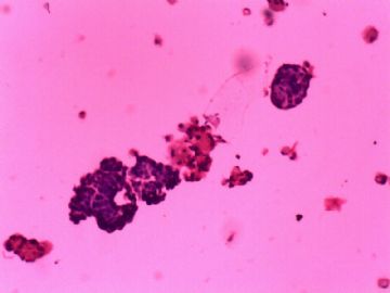
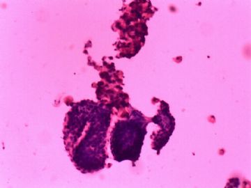
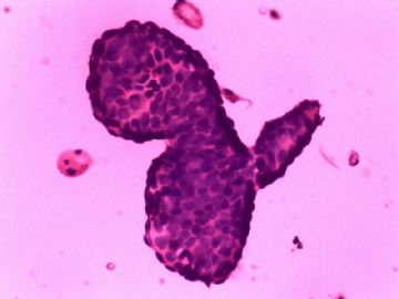
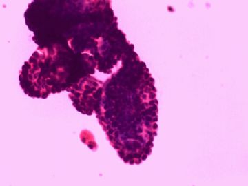
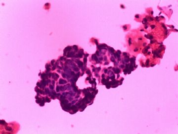
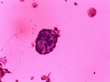
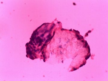
- 大家看看这一例乳腺印片够癌吗?
非常感谢陈博士的精彩 的点评,给了我们很多学习的地方.
在乳头溢液的印片涂片中,诊断癌确实风险性太大,没有必要把这个风险主动转交到自己头上.
陈博士说的,也是我们在这一例病例上工作的小失误,我们应该提一下,建议临床活检或是手术做冰冻.
感谢陈博士的教诲!
但这一例病例因为我没有去摸肿块,所以不知道病人是否有肿块.不过我曾经遇到过这样一例病例,也是乳头的溢液涂片,图像和这例差不多(没有这么典型,不过也有呈乳头状的异型腺上皮),当时我摸了病人的双侧乳腺和腋窝都没有肿块.
请教陈博士,遇到这种情况我们应该如何处理呢?
OK! If this case is cytology of nipple discharge, my diagnosis would be "atypical epithelial cells, favor an origin from a papillary neoplasm". I would recommend biopsy if clinically indicated. Please note the following points:
1) Cytology itself is very difficult to distinguish between a papilloma vs. a low-grade papillary carcinoma, thus I used term papillary neoplasm
2) The fact that your surgical specimen showed carcinoma does not mean that you can diagnose carcinoma on cytology. Remember that the difference between cytology and histology is that cytology has both diagnostic and screening functions. In those cases that cytology have difficulties, then it mainly serves as a screening tool (such as Pap test), then the "so called Gold-standard" will be the patient get proper management. With my above diagnosis, the patient will get the right management.
I have to apologize for my limited knowledge. What are these slides? Are they from FNA or touch Prep?
In either case, I would be very cautious to diagnose carcinoma. The slides are in bad quality and it appears that there are some papillary groups of epithelial cells, that's all I can see and say at this moment.
I strongly oppose the diagnostic terminology of "导管内乳头状瘤", on cytology, you can NOT see tissue invasion, so you can not tell if it is in-situ carcinoma or invasive carcinoma. Just like in Pap test, you do NOT diagnose "micro-invasive carcinoma".

