| 图片: | |
|---|---|
| 名称: | |
| 描述: | |
- B1760Breast LCIS involving sclerosing adenosis+ADH (cqz 11)
| 姓 名: | ××× | 性别: | 年龄: | ||
| 标本名称: | |||||
| 简要病史: | |||||
| 肉眼检查: | |||||
I assume that all of you still are enjoying your New Year Holiday. Send here a case for your consideration.
F/50y Breast core biopsy (5 cores)
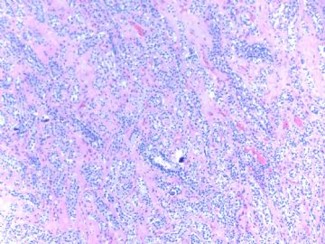
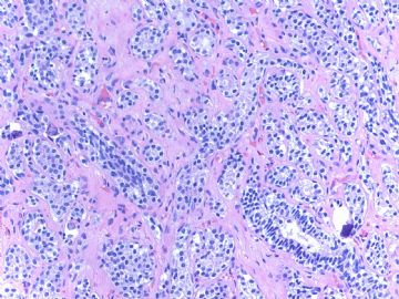
Lesion 1
Fig 1 10x
Fig 2 20x
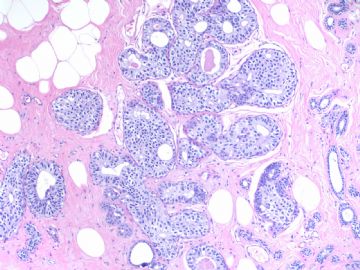
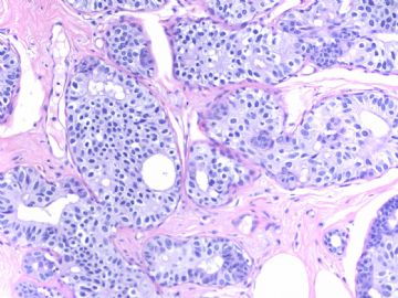
Lesion 2
fig 3 10x
Fig 4 20x
-
本帖最后由 于 2009-02-22 10:46:00 编辑
相关帖子
- • 乳腺肿物
- • 导管原位癌or不典型增生
- • 乳腺癌?
- • 女性 冰冻为乳腺浸润性导管癌,现切除标本,肿块旁组织
- • 女性 33岁 乳腺肿块
- • 乳腺包块
- • 乳腺两个相邻导管内的病变
- • 乳腺肿物
- • 乳腺包块。33岁
- • 左乳肿块,协助诊断
-
本帖最后由 于 2009-02-02 23:52:00 编辑
Dr. Zhao的意思,大家以后可以直接称呼Zhao或者Dr. Zhao
这个病例很有趣,鉴别范围很广泛(不许笑话我,呵呵),包括UDH,ADH,IDC
因为不存在小叶结构,图1和图2中硬化性背景上那些模糊的小管结构和近似实性的条索,需要排除浸润癌(尽管不太像,因为可以辨认肌上皮细胞,可是……小心一些没坏处,呵呵),可以染肌上皮标记。
这例UDH与ADH的区分对我来说真的太困难了。似乎皆有(我汗,再次强调,不许笑话我)。是否还有可能ALH/LCIS累犯小叶,E-ca和p120有帮助。
期待Dr.Zhao解惑,谢谢!

华夏病理/粉蓝医疗
为基层医院病理科提供全面解决方案,
努力让人人享有便捷准确可靠的病理诊断服务。
Hi, Dr. Zhao,
Happy Spring Festival!
In my opinion, both are at least atypical hyperplasia(case 1 lobular and case 2 dutal?). carcinoma should be futher ruled out with p63 CK5/6 immunostaining. Note the micro-calcification in case 1, which is implying "bad lesion".
Looking forward to the final diagnosis and explanation.
Many thanks!

- If you have great talents, industry will improve them; if you have but moderate abilities, industry will supply their deficiency. 如果你很有天赋,勤勉会使其更加完美;如果你能力一般,勤勉会补足其缺陷。
-
本帖最后由 于 2009-01-31 17:23:00 编辑
Ok! Dr.zhao: You are right. I was happy to read your words. I will call you Dr.zhao, you can call me “望月”too. Your guess is correct. i am a lady ,Because I like the moon, in the quiet of the night, often looking at the moon reverie......hehe
 ,My name is wangyue(望月)。
- 广州金域病理
-
本帖最后由 于 2009-01-31 03:53:00 编辑
Dr. 天山望月 :
Internet is interesting. I even do not know you are male or female. I guess you may be a female because you work hard and study carefully. These features often are characteristic of female. Men should not feel angery because I am male also.
I like the way of your analysis.It does not mean they are right or wrong. I do not want to influence other people's oppinion.
好像看的越多,考虑的越多,越不敢猛下诊断了. It means that you become more and more expert. I choose some cases and send here with some difficulties. It is not easy to make dx by H&E only. You should think over.
About the call for people: In the US you can call the first name for your boss or your professor. In hospitals generally people call physicians--Dr. Wang, Li, Zhang et al. Even you meet world famous doctors, you stil call Dr. xxx. For friends among physicians you can call first name. In China, people use teachers too often. I favor that your guy call me zhao or Dr. Zhao, but not teacher zhao. I feel more comfortable for zhao or dr. zhao. I can call you name or Dr. xx
谢谢赵老师!春节假期非常开心分享您的病例!
我觉得:1、鉴别小叶和导管病变,E-Ca和P120。2、鉴别UDH、ADH、DCIS或小叶病变累及导管、浸润癌标记:ER, PR, CK5/6,P63,SMMH.3、需鉴别肌上皮病变。
Lesion 1纤维化组织穿插在腺泡、导管间,一些导管上皮单一,异型,圆形细胞,胞浆有些透亮,可见钙化。纤维组织中有单个大异型细胞(需排除小叶病变),一些管腔周围有肌上皮和基底膜。
Lesion 2管腔内上皮不太单一,但有异型,可见核仁,边窗有些涨大,也有些细胞流水样排列。
请赵老师赐教!再次感谢!
(注:好像看的越多,考虑的越多,越不敢猛下诊断了 ,还是功夫不到啊,期待赵老师多出练习和考题了,相信功夫不负有心人!)

- 广州金域病理
