| 图片: | |
|---|---|
| 名称: | |
| 描述: | |
- B1757Invasive ductal ca look as DCIS-the importance of myoepithelial marker (cqz-10)
| 姓 名: | ××× | 性别: | 年龄: | ||
| 标本名称: | |||||
| 简要病史: | |||||
| 肉眼检查: | |||||
We are in 2009 already. I send this easy case for you.
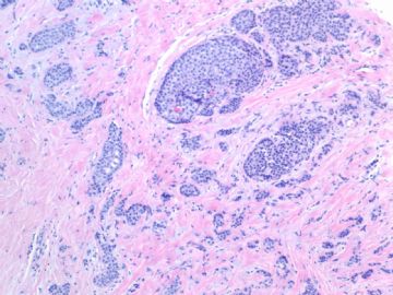
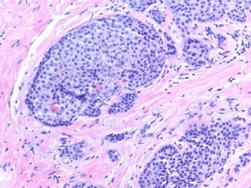
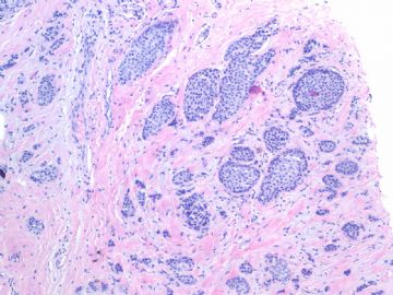
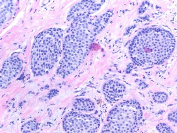
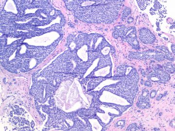
Breast core biopsy:
Fig 1, 2: one area 100x, 200x
Fig 3, 4: the second area 100x, 200x
Fig 5: the third area 100x
What is your interpretation?
标签:DCIS 乳腺浸润性导管癌
-
本帖最后由 于 2009-07-19 05:14:00 编辑
相关帖子
- • 乳腺肿物
- • 乳腺肿物
- • 乳腺癌吗??
- • 左乳肿块,协助诊断
- • 乳腺肿物
- • 乳腺小管癌?
- • 左乳肿瘤--浸润性导管癌?
- • 看看这是那个类型的乳腺癌?
- • 乳腺肿物,请大家帮忙会诊是恶性的吗??
- • 急`1`1`1`1乳腺肿物,请大家帮忙会诊
×参考诊断
-
本帖最后由 于 2009-01-24 12:20:00 编辑
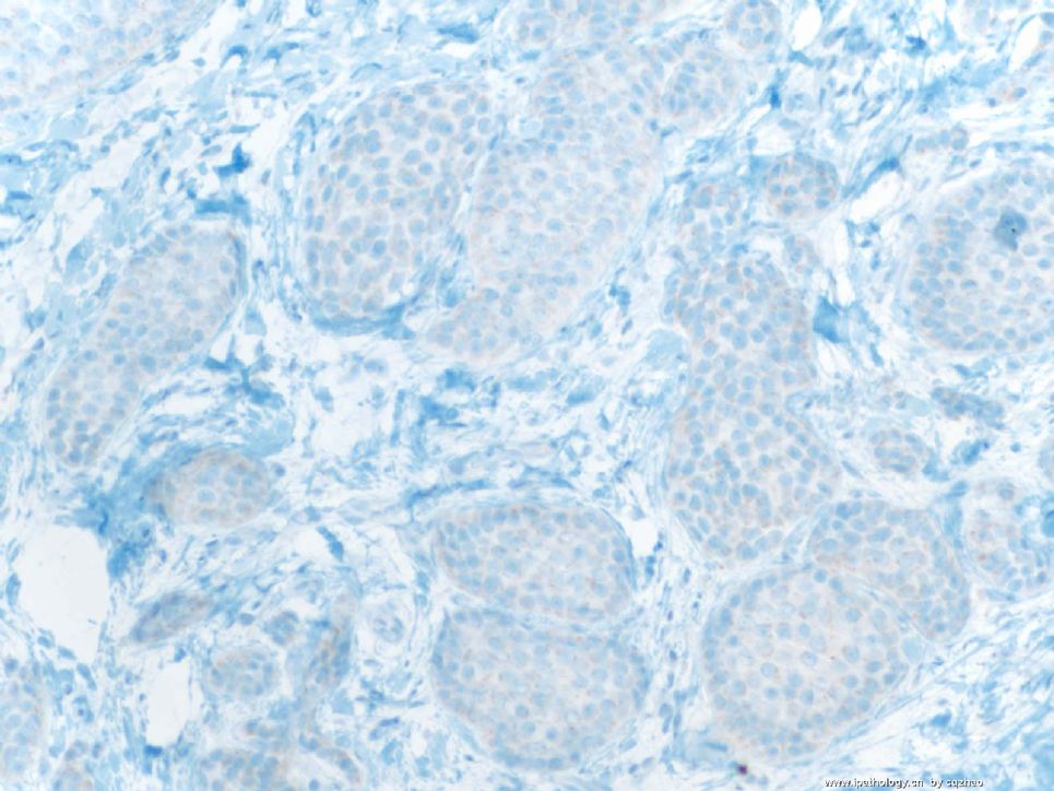
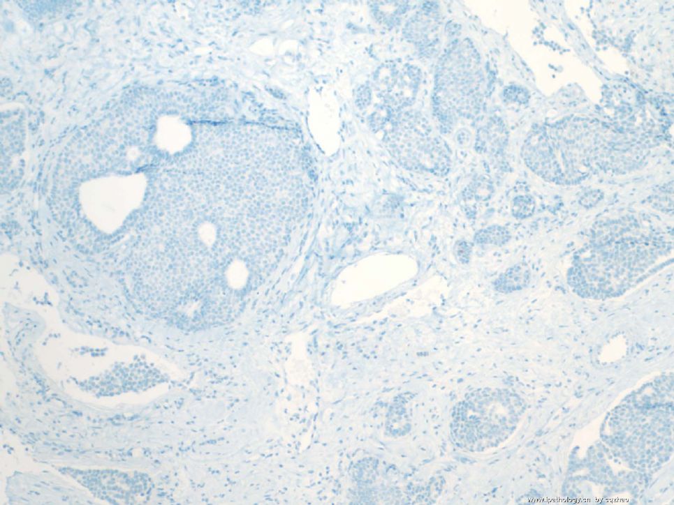
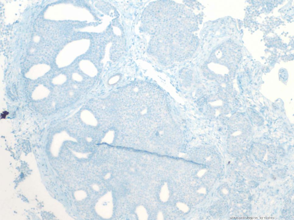
P63 stains
Fig 1 first area
Fig 2 second area with SMMHC positive-looking.
Fig 3 second area. I repeat P63 stain
Now I want to see your interpretation.
I chose some cases, sent here, and tried to demonstrate some points for each case.
For this case, we are sure that invasive carcinoma is present. Now the question is if DCIS is present or all are invasive carcinoma.
You guys can think about it.
名称:图1
描述:图1
名称:图2
描述:图2
名称:图3
描述:图3
