| 图片: | |
|---|---|
| 名称: | |
| 描述: | |
- B1757Invasive ductal ca look as DCIS-the importance of myoepithelial marker (cqz-10)
| 姓 名: | ××× | 性别: | 年龄: | ||
| 标本名称: | |||||
| 简要病史: | |||||
| 肉眼检查: | |||||
We are in 2009 already. I send this easy case for you.
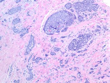
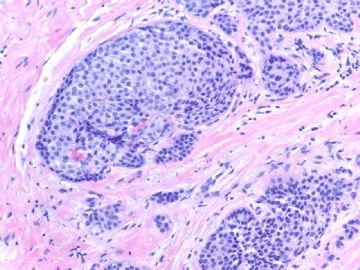
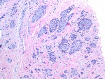
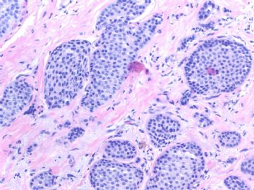
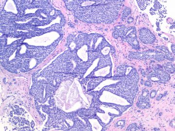
Breast core biopsy:
Fig 1, 2: one area 100x, 200x
Fig 3, 4: the second area 100x, 200x
Fig 5: the third area 100x
What is your interpretation?
-
本帖最后由 于 2009-07-19 05:14:00 编辑
相关帖子
- • 乳腺肿物
- • 乳腺肿物
- • 乳腺癌吗??
- • 左乳肿块,协助诊断
- • 乳腺肿物
- • 乳腺小管癌?
- • 左乳肿瘤--浸润性导管癌?
- • 看看这是那个类型的乳腺癌?
- • 乳腺肿物,请大家帮忙会诊是恶性的吗??
- • 急`1`1`1`1乳腺肿物,请大家帮忙会诊
Happy new year, Dr.cqzhao!
These pictures show LCIS or DCIS, which can be differentiated by each other for immunostaining of p120 and E-cadherin. In the stromal pseudoinvasion is presented, and p63 immunostaining may confirm it.

华夏病理/粉蓝医疗
为基层医院病理科提供全面解决方案,
努力让人人享有便捷准确可靠的病理诊断服务。
-
i feel it has both LCIS (first several photos) and DCIS (last photo), low nuclear grade. Invasive cancer cannot be excluded because the first several photos show small clusters of neoplastic cells in desmoplastic-like stroma. E-cadherin and p63 will certainly help.
-
Happy New Year Dr.zhao, Thank you for sharing interesting breast cases.It's very nice to learn your cases.
I think the case has both LCIS and DCIS, invasive lobular carcinoma can not be excluded. Looking forward for the your explanation and immunostaining results.
-
stevenshen 离线
- 帖子:343
- 粉蓝豆:2
- 经验:343
- 注册时间:2008-06-03
- 加关注 | 发消息
-
本帖最后由 于 2009-01-19 12:41:00 编辑
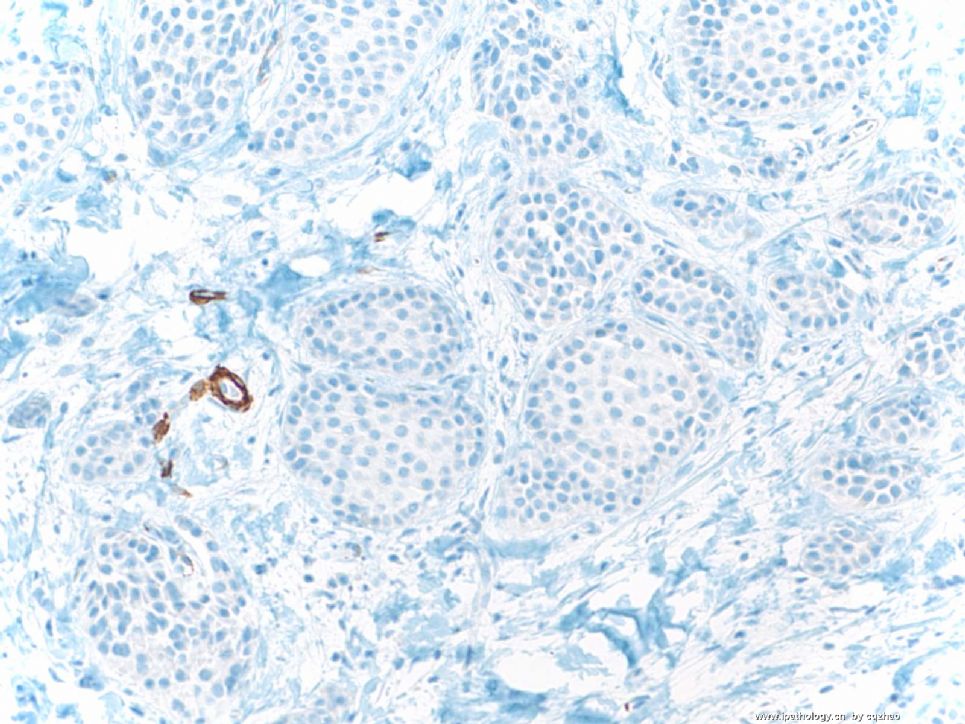
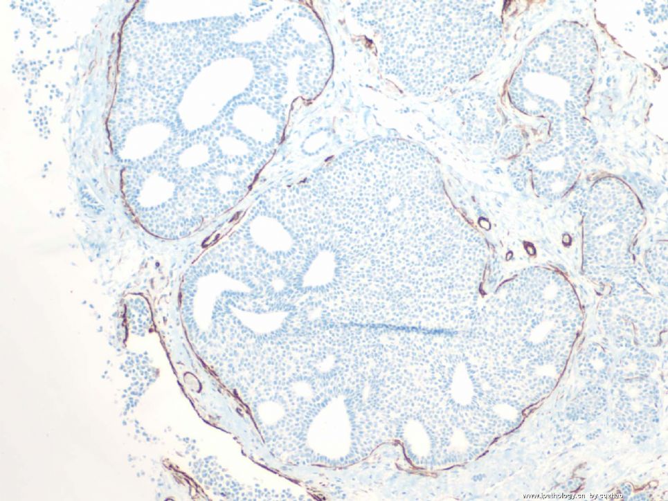
IHC: smooth muscle myosin heavy chain
Fisrt Fig. the area for first several figs of H&E
Second Fig. the area for the last photo of H&E.
I am a little surprise to read the response. Now what is your final diagnosis for this case?
名称:图1
描述:图1
名称:图2
描述:图2
-
本帖最后由 于 2009-01-24 12:20:00 编辑
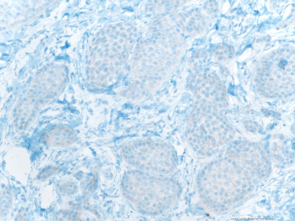
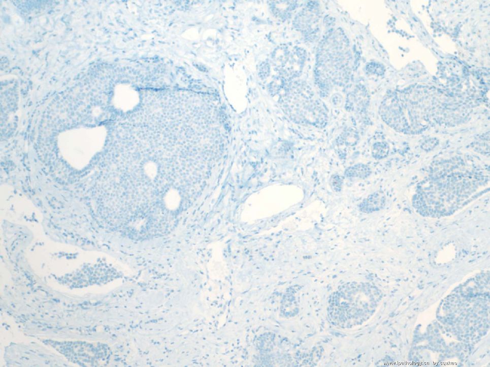
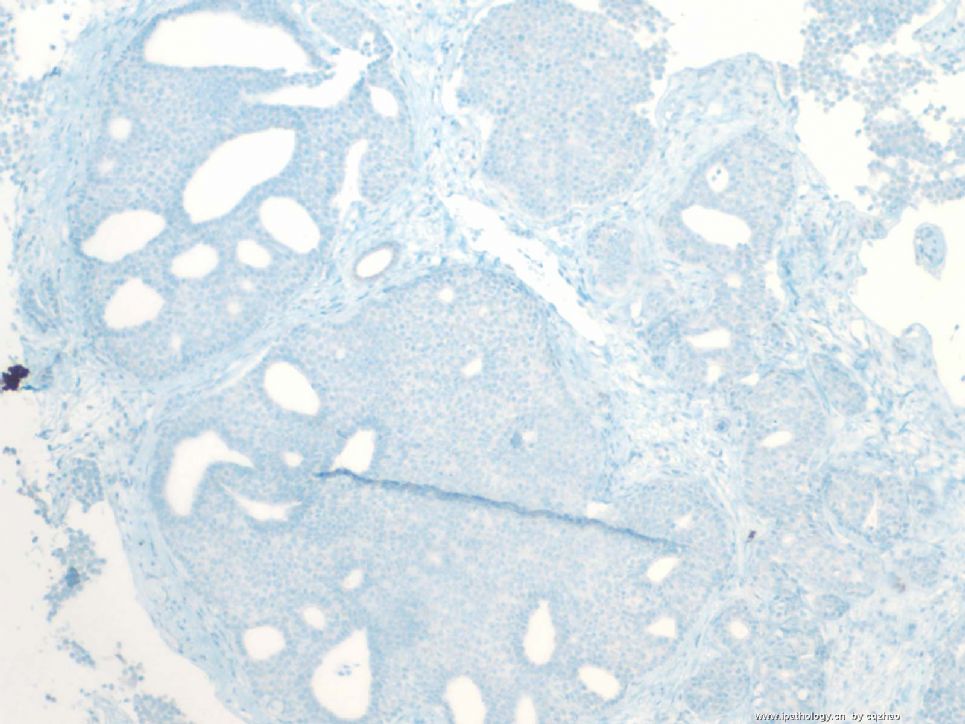
P63 stains
Fig 1 first area
Fig 2 second area with SMMHC positive-looking.
Fig 3 second area. I repeat P63 stain
Now I want to see your interpretation.
I chose some cases, sent here, and tried to demonstrate some points for each case.
For this case, we are sure that invasive carcinoma is present. Now the question is if DCIS is present or all are invasive carcinoma.
You guys can think about it.
名称:图1
描述:图1
名称:图2
描述:图2
名称:图3
描述:图3


 又猜不透赵老师的心思了,可否给一点方向呢?


