共3页/45条首页上一页123下一页尾页
回复:46 阅读:7409
罕见病例!!!

zhangjianxin 离线

帖子:111
粉蓝豆:11
经验:111
注册时间:2007-03-06
加关注  |  发消息
楼主 发表于 2008-12-24 17:04|举报|关注(9)
浏览排序[ 顺序 逆序 楼主 支持 精彩 ]  快捷回复

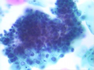
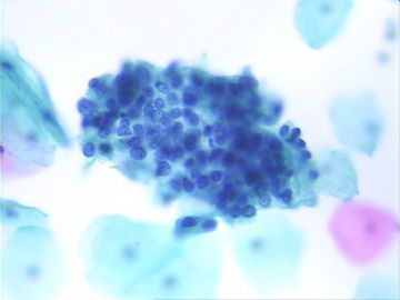
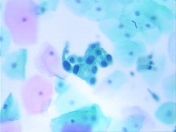
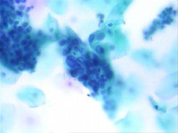
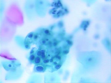
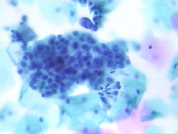
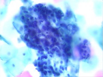
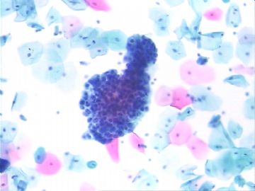
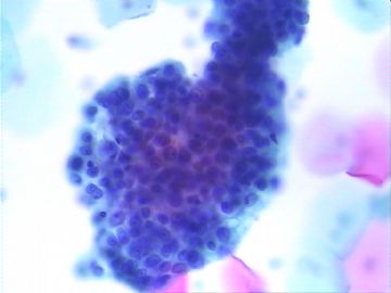
33岁,月经紊乱,不规则出血史。该病例在宫颈细胞学较为罕见,请大家欣赏!

  • 罕见病例!!!图1
    图1
  • 罕见病例!!!图2
    图2
  • 罕见病例!!!图3
    图3
  • 罕见病例!!!图4
    图4
  • 罕见病例!!!图5
    图5
  • 罕见病例!!!图6
    图6
  • 罕见病例!!!图7
    图7
  • 罕见病例!!!图8
    图8
  • 罕见病例!!!图9
    图9
  • 罕见病例!!!图10
    图10
标签:
0
添加参考诊断
×参考诊断
  

cqzhao 离线

帖子:5518
粉蓝豆:1071
经验:5755
注册时间:2008-09-29
加关注  |  发消息
21 楼    发表于2009-01-08 06:09:00举报|引用
返回顶部 | 快捷回复
 I remember this case and come here to check the result. I like above discussion. We all have equal power and right to mention our oppinions here. We will not take any responsbility for what we said. For the true case we are not wrong if we report AGC. However we have to contact with patient's clinician to make sure the patient will have cervical and endometrial sampling. It is true that we must be very cautious to call carcinoma in Pap for these young patients.
0
回复

cqzhao 离线

帖子:5518
粉蓝豆:1071
经验:5755
注册时间:2008-09-29
加关注  |  发消息
22 楼    发表于2009-01-08 06:22:00举报|引用
返回顶部 | 快捷回复

 Read photos again. generally speaking , clear cell carcinomas have more ugly cytology, but these cells are relative bland even though we can appreciate the pleomorphism of cells. So endometrioid carcinoma???

zhangjianxin 

Thank for the interesting case. We are waiting for your surgical results.

Do you have residule fluid? You can ask Lab to make a cell block if you have. It may be helpful. My feeling is there are many clusters of cells in the fluid.

0
回复

兰青风采 离线

帖子:101
粉蓝豆:1
经验:101
注册时间:2008-07-13
加关注  |  发消息
23 楼    发表于2009-01-08 13:01:00举报|引用
返回顶部 | 快捷回复

等待中。。。。。。

0
回复

赌具 离线

帖子:380
粉蓝豆:12
经验:801
注册时间:2008-03-06
加关注  |  发消息
24 楼    发表于2008-12-24 19:03:00举报|引用
返回顶部 | 快捷回复
 腺癌,不除外腺样鳞癌?
0
回复
signature
平凡一人, 风顺一帆, 平淡一生.

海浪信使 离线

帖子:1547
粉蓝豆:161
经验:2339
注册时间:2006-09-26
加关注  |  发消息
25 楼    发表于2008-12-24 19:37:00举报|引用
返回顶部 | 快捷回复
本帖最后由 于 2008-12-24 19:53:00 编辑  宫颈腺癌?
0
回复
signature
当你有选择的时候,不是选择正确的,而是选择不让你后悔的!

yunhuixin 离线

帖子:13
粉蓝豆:1
经验:13
注册时间:2008-12-24
加关注  |  发消息
26 楼    发表于2008-12-24 19:58:00举报|引用
返回顶部 | 快捷回复
 核大,深染,可见核仁,至少是高度核异质细胞.
0
回复

thlcp 离线

帖子:911
粉蓝豆:6
经验:911
注册时间:2008-10-26
加关注  |  发消息
27 楼    发表于2008-12-24 20:17:00举报|引用
返回顶部 | 快捷回复
 腺鳞癌
0
回复

houfang 离线

帖子:116
粉蓝豆:0
经验:120
注册时间:2008-07-04
加关注  |  发消息
28 楼    发表于2008-12-24 20:47:00举报|引用
返回顶部 | 快捷回复
复习宫颈腺癌的主要特征:
1、癌细胞排列呈单个散在或双层片状或团状三维结构;
2、胞核增大,核膜增厚而不规则;
3、核仁明显或增大或多核仁;
4、胞质呈蓝染,也可红染,大小不等或囊性空泡出现
此病例符合以上特点。
0
回复

故乡 离线

帖子:703
粉蓝豆:318
经验:961
注册时间:2007-12-15
加关注  |  发消息
29 楼    发表于2008-12-24 21:30:00举报|引用
返回顶部 | 快捷回复
 同意腺癌
0
回复

法师 离线

帖子:1077
粉蓝豆:14
经验:1077
注册时间:2007-02-06
加关注  |  发消息
30 楼    发表于2008-12-24 21:51:00举报|引用
返回顶部 | 快捷回复
以下是引用thlcp在2008-12-24 20:17:00的发言:

 腺鳞癌

0
回复

天职 离线

帖子:111
粉蓝豆:1
经验:111
注册时间:2008-10-12
加关注  |  发消息
31 楼    发表于2008-12-25 18:30:00举报|引用
返回顶部 | 快捷回复
 好文章
0
回复

huijunren 离线

帖子:4
粉蓝豆:5
经验:4
注册时间:2018-12-12
加关注  |  发消息
45 楼    发表于2018-12-12 17:43:08举报|引用
来自Android 返回顶部 | 快捷回复
最终病理结果是什么呢?
0
回复

悟道(迷) 离线

帖子:63
粉蓝豆:1586
经验:460
注册时间:2014-06-26
加关注  |  发消息
40 楼    发表于2017-02-13 10:19:35举报|引用
返回顶部 | 快捷回复

最终结果还没有解开


0
回复
signature
天道酬勤 强者生存

zhangjianxin 离线

帖子:111
粉蓝豆:11
经验:111
注册时间:2007-03-06
加关注  |  发消息
34 楼    发表于2009-01-02 18:10:00举报|引用
返回顶部 | 快捷回复

        该病例比较特殊,希望大家参与讨论!我觉得华夏病理网给我们这么好的交流平台,就是希望大家在平时忙碌得工作中,遇到的特殊病理拿出来一块交流,共同提高。

        该病例之所以特殊:1、细胞之间的结构较为松散。2、部分细胞胞浆比较丰富。3、部分细胞有透亮的胞浆。所以我觉得该病例的分型值得我们推敲!

0
回复

wangdingding 离线

帖子:1474
粉蓝豆:98
经验:6042
注册时间:2006-10-19
加关注  |  发消息
35 楼    发表于2009-01-02 18:42:00举报|引用
返回顶部 | 快捷回复
 倾向低分化鳞癌
0
回复

有福不在忙 离线

帖子:2161
粉蓝豆:42
经验:3448
注册时间:2008-11-22
加关注  |  发消息
36 楼    发表于2009-01-02 18:46:00举报|引用
返回顶部 | 快捷回复
 我也倾向腺癌,但要排除滋养细胞(肿瘤)。
0
回复
signature
有福不在忙

小荷 离线

帖子:8546
粉蓝豆:97
经验:12725
注册时间:2006-09-11
加关注  |  发消息
37 楼    发表于2009-01-03 01:18:00举报|引用
返回顶部 | 快捷回复
 腺癌,转移的?
0
回复
signature
没有完美的个人,只有完美的团队

cqzhao 离线

帖子:5518
粉蓝豆:1071
经验:5755
注册时间:2008-09-29
加关注  |  发消息
38 楼    发表于2009-01-03 02:17:00举报|引用
返回顶部 | 快捷回复
本帖最后由 于 2009-01-03 02:54:00 编辑

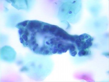
Cellular specimen with cluster highly atypical cells showing pleomorphism, prominnat nuclei, and abundant cytoplasm with clear appearance in some cells.

Agree with above. It looks like adenocarcinoma. It  can be endocercial or endometrial origins or metastatic tumor.

Tumor type can be clear cell carcinoma or other. Based on age and cytology, It can be endometrial endometrioid carcinoma. secretory variant. Remeber that I say "can" be....

Anyway in the Pap test it is good enough if you report adenocarcinoma or AGC favor neoplasma if you want to leave some space for you. For this case, it may provde you more information if you can prepare a cell block or do some IHC if you have a good cell block.

I mentioned here many times that Pap test is a screening test. We should keep this priciple in our mind when we evaluate Pap test.

Thank for the excellent case and photos. Waiting for your final histologic results.
0
回复

liuqingle 离线

帖子:204
粉蓝豆:25
经验:217
注册时间:2007-08-14
加关注  |  发消息
39 楼    发表于2009-01-03 08:14:00举报|引用
返回顶部 | 快捷回复
 
  腺鳞癌

0
回复

zhangjianxin 离线

帖子:111
粉蓝豆:11
经验:111
注册时间:2007-03-06
加关注  |  发消息
40 楼    发表于2009-01-06 15:51:00举报|引用
返回顶部 | 快捷回复
 今天电话随访,因患者经期刚结束,还未去医院就诊。有结果会及时发上来!
0
回复
回复:46 阅读:7409
共3页/45条首页上一页123下一页尾页
【免责声明】讨论内容仅作学术交流之用,不作为诊疗依据,由此而引起的法律问题作者及本站不承担任何责任。
快速回复
进入高级回复
您最多可输入10000个汉字,按 "Ctrl" + "Enter" 直接发送
搜索回复/乘电梯 ×
按内容
按会员
乘电梯
合作伙伴
友情链接