共8页/150条首页上一页45678下一页尾页
回复:150 阅读:16938
an easy case for you-Pap test (cqz 3)

cqzhao 离线

帖子:5518
粉蓝豆:1071
经验:5755
注册时间:2008-09-29
加关注  |  发消息
楼主 发表于 2008-12-12 11:42|举报|关注(0)
浏览排序[ 顺序 逆序 楼主 支持 精彩 ]  快捷回复

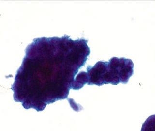
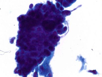
Pap smears are too difficult, especially for photos in computer. Give you an easy one for fun.

A women aged over 50

How will you sign out the case?

  • an easy case for you-Pap test  (cqz 3)图1
    图1
  • an easy case for you-Pap test  (cqz 3)图2
    图2
标签:
0
添加参考诊断
×参考诊断
  

天山望月 离线

帖子:4902
粉蓝豆:83
经验:5092
注册时间:2007-02-03
加关注  |  发消息
101 楼    发表于2009-01-03 21:02:00举报|引用
返回顶部 | 快捷回复
 反复看细胞块图,心里总觉得不踏实,不排除癌变,要了解一下B超检查宫腔情况,劲管情况,分段诊刮。
0
回复
signature
广州金域病理

cqzhao 离线

帖子:5518
粉蓝豆:1071
经验:5755
注册时间:2008-09-29
加关注  |  发消息
102 楼    发表于2009-01-03 21:53:00举报|引用
返回顶部 | 快捷回复
以下是引用天山望月在2009-1-3 20:40:00的发言:

 想悄悄问问赵老师:做细胞块,如果病变明确,是直接诊断呢,还是先联系临床,再进一步工作后诊断?

做细胞块收费吗?

Cell block and Pap test are from the same sample. If we are not sure in the Pap test but we are sure it is maligancy in cell block. We still can call mailignancy. It is one diagnosis. It is the same as we make dx for FNA.Our dx is based smears and cell blocks. We do not need to contact clinicians before we do cell block. We do not do cell blocks often, just only for some special cases. I do not think we charge patients. Generally I cantact cyto lab in person and ask cytotechnitions do a favor. Of cause you can charge patients.
0
回复

cqzhao 离线

帖子:5518
粉蓝豆:1071
经验:5755
注册时间:2008-09-29
加关注  |  发消息
103 楼    发表于2009-01-03 21:57:00举报|引用
返回顶部 | 快捷回复
 Now the question is that  it is carcinoma or not. if it is carcinoma it is from endometrial or endocervical origins. We have cell block and can do IHC. Which ihc is useful for dx of malignancy?
0
回复

天山望月 离线

帖子:4902
粉蓝豆:83
经验:5092
注册时间:2007-02-03
加关注  |  发消息
104 楼    发表于2009-01-03 22:07:00举报|引用
返回顶部 | 快捷回复
 Thank Dr.zhao! i  understand.
0
回复
signature
广州金域病理

有福不在忙 离线

帖子:2161
粉蓝豆:42
经验:3448
注册时间:2008-11-22
加关注  |  发消息
105 楼    发表于2009-01-03 22:10:00举报|引用
返回顶部 | 快捷回复
 我看像是恶性,CEA和Vimentin对鉴别内膜和宫颈来愿有帮助,宫颈腺癌CEA+,Vimentin-;内膜癌相反。
0
回复
signature
有福不在忙

天山望月 离线

帖子:4902
粉蓝豆:83
经验:5092
注册时间:2007-02-03
加关注  |  发消息
106 楼    发表于2009-01-03 22:54:00举报|引用
返回顶部 | 快捷回复

 回复104楼:

如果有活检标本,可以观察子宫内膜癌或宫颈腺癌各自的癌前病变,看过度形态,有利于鉴别,

IHC有帮助:宫内膜癌:CEA±(50%),VIM+,CAM5.2+,宫颈腺癌CEA+(100%),VIM-,CAM5.2-.

期待最后结果。。。

不简单的病例,锻炼思维,磨练意志!

0
回复
signature
广州金域病理

小荷 离线

帖子:8546
粉蓝豆:97
经验:12725
注册时间:2006-09-11
加关注  |  发消息
107 楼    发表于2009-01-03 23:14:00举报|引用
返回顶部 | 快捷回复
 哇塞!继续
0
回复
signature
没有完美的个人,只有完美的团队

cqzhao 离线

帖子:5518
粉蓝豆:1071
经验:5755
注册时间:2008-09-29
加关注  |  发消息
108 楼    发表于2009-01-05 09:47:00举报|引用
返回顶部 | 快捷回复
本帖最后由 于 2009-01-05 09:54:00 编辑

 Fig:  cell block, Ki67 stain.

Happy to see some of you had suggestion about IHC for the origins. Now forget the origins, just think how you will sign the report based the Pap, cell block, and ki67.

AGC

AGC, favor neoplastic

Adenocarcinoma.

choose one from above three

名称:图1
描述:图1
0
回复

天山望月 离线

帖子:4902
粉蓝豆:83
经验:5092
注册时间:2007-02-03
加关注  |  发消息
109 楼    发表于2009-01-05 13:12:00举报|引用
返回顶部 | 快捷回复

 IHC:Ki67很高的(  >80%),结合HE乳头状形态,我选Adenocarcinoma.

赵老师:此例分段诊刮了吗?期待欣赏哦!

0
回复
signature
广州金域病理

天山望月 离线

帖子:4902
粉蓝豆:83
经验:5092
注册时间:2007-02-03
加关注  |  发消息
110 楼    发表于2009-01-05 13:20:00举报|引用
返回顶部 | 快捷回复
 再回顾81楼病例,真是太玄了,单从细胞片,如果没有进一步工作,仅提示病人1个月后复查,将会延误肿瘤治疗。因此,细胞片上,只要有一点蛛丝马迹,都要慎之又慎,多做工作,结合临床病史。
0
回复
signature
广州金域病理

兰青风采 离线

帖子:101
粉蓝豆:1
经验:101
注册时间:2008-07-13
加关注  |  发消息
111 楼    发表于2009-01-06 21:35:00举报|引用
返回顶部 | 快捷回复

 谢谢赵老师,在细胞网里这个贴子没有坚持下去;因为我也没有时间经常上网;但是只要有赵老师的回帖我都得认真学习,今天把整个看完之后;感觉我只是一个连井底的青蛙都不是。刚开始看宫颈细胞学以为很简单;越看感觉越看不懂,也越来越小心;最后以至于不敢报,对自己产生怀疑;一直想找一位名师指点,终于见到赵老师的执着和渊博,真诚的说一声谢谢;也祝福我们这里的所有同仁们在我们传统节日来之前开开心心,健康过大年。

     这个病例在如果只有前面细胞图片,我会选择报AGC,如果要我签发。如果不签发我当时在细胞网“忽悠”的是AIS;最后结合细胞块和Ki-67结果,我只能选择Adenocarcinoma了,谢谢!

0
回复

小荷 离线

帖子:8546
粉蓝豆:97
经验:12725
注册时间:2006-09-11
加关注  |  发消息
112 楼    发表于2009-01-06 22:46:00举报|引用
返回顶部 | 快捷回复
 欢迎新朋友
0
回复
signature
没有完美的个人,只有完美的团队

cqzhao 离线

帖子:5518
粉蓝豆:1071
经验:5755
注册时间:2008-09-29
加关注  |  发消息
113 楼    发表于2009-01-07 01:12:00举报|引用
返回顶部 | 快捷回复

  To 兰青风采:

Glad to see you here.  I like to read your discussion. Every one is the same on line. No one has any responsbility for patient care here. It is a good place for discussion, especialy for people who really want to learn sth.

Welcome you here and wish you can join in the discussion activily if you have additional time.

0
回复

cqzhao 离线

帖子:5518
粉蓝豆:1071
经验:5755
注册时间:2008-09-29
加关注  |  发消息
114 楼    发表于2009-01-07 02:05:00举报|引用
返回顶部 | 快捷回复

 The Pap smear demonstrates clusters of hyperchromatic crowded groups of cells. Based on the Pap only we should call AGC at least. It is ok If you call AGC because women should get biopsy. It is not a good call if we call negative or reactive. The causes of AGC are lesions. They range from completely normal, to hyperplastic, to neoplastic conditions.  If you call reacitve it means that you definitely think it cannot be hyperplastic or neoplastic conditions. Agree with all of your interpretation. The Pap was singed out adenocarcinoma based on Pap, cell block, and IHC.

0
回复

cqzhao 离线

帖子:5518
粉蓝豆:1071
经验:5755
注册时间:2008-09-29
加关注  |  发消息
115 楼    发表于2009-01-07 02:06:00举报|引用
返回顶部 | 快捷回复
本帖最后由 于 2009-01-07 02:22:00 编辑

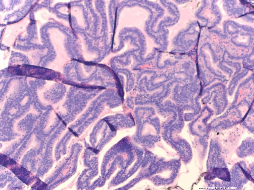
The women had cervical biopsy.

both 100x

Now we agree that it is an adenocarcinoma case. Let us discuss the origins of the tumor, endocervical or endometrial. As gynecologic pathologists we oftern meet this situation. Gynecologists hope to know the origin of the adenocarcinoma when we report adenocarcinoma in cervical biopsy report. They wish to know it is endocervical origin or endometrial carcinoma metastatic to endocervix or other tumors from location to endocervix. Often we cannot answer the question even thpugh we perform many IHC studies.

Anyway, what IHC would you ordered if it is your case? What is your guess (I said guess, not diagnosis. So every one can give a guess) of the origin based on the H&E?

Now we transfer the Pap cytology to gynecologic surgical pathology.


名称:图1
描述:图1
名称:图2
描述:图2
0
回复

兰青风采 离线

帖子:101
粉蓝豆:1
经验:101
注册时间:2008-07-13
加关注  |  发消息
116 楼    发表于2009-01-08 20:39:00举报|引用
返回顶部 | 快捷回复
 这个来源从HE的片子来说还真困难,至少我很难判断是来源于宫颈管的内膜样腺癌还是宫腔的内膜腺癌;在临床医生分段诊刮的组织取材位置对我们判断来源或许有帮助;如果两者都有癌组织的话,到底是哪个是原发灶那还真是一个“迷”;也许还有其他的手段能证明,但是我确实不懂,请赵老师指点。谢谢!
0
回复

cqzhao 离线

帖子:5518
粉蓝豆:1071
经验:5755
注册时间:2008-09-29
加关注  |  发消息
117 楼    发表于2009-01-10 07:36:00举报|引用
返回顶部 | 快捷回复
本帖最后由 于 2009-01-10 07:40:00 编辑

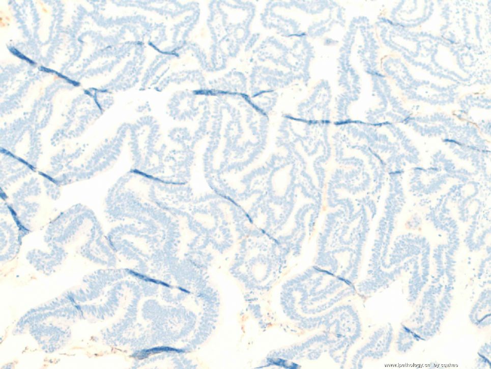
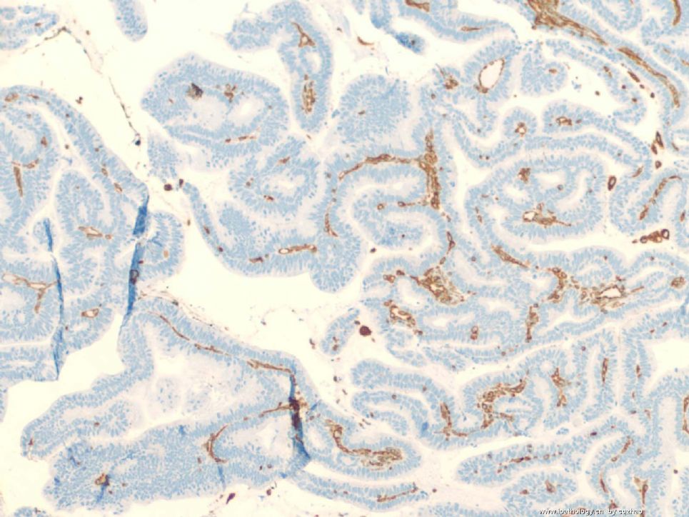
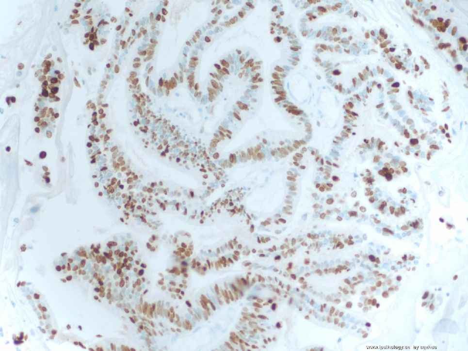
 IHC result of above biopsy

f1 CEA

f2 ER

F3 P16

F4 Vimentin

We have to sign out the case and no more investigation.

How will you sign out the case of cervical biopsy?


名称:图1
描述:图1

名称:图2
描述:图2

名称:图3
描述:图3

名称:图4
描述:图4
0
回复

xiaogang 离线

帖子:5664
粉蓝豆:48
经验:6068
注册时间:2008-12-18
加关注  |  发消息
118 楼    发表于2009-01-10 11:09:00举报|引用
返回顶部 | 快捷回复
 疑似腺癌,建议活检。
0
回复

xiaogang 离线

帖子:5664
粉蓝豆:48
经验:6068
注册时间:2008-12-18
加关注  |  发消息
119 楼    发表于2009-01-10 11:14:00举报|引用
返回顶部 | 快捷回复
 看到病理片报腺癌无疑。
0
回复

cqzhao 离线

帖子:5518
粉蓝豆:1071
经验:5755
注册时间:2008-09-29
加关注  |  发消息
120 楼    发表于2009-01-10 20:55:00举报|引用
返回顶部 | 快捷回复
以下是引用xiaogang在2009-1-10 11:14:00的发言:

 看到病理片报腺癌无疑。

what type? possible origin beased on morphology and IHC.
0
回复
回复:150 阅读:16938
共8页/150条首页上一页45678下一页尾页
【免责声明】讨论内容仅作学术交流之用,不作为诊疗依据,由此而引起的法律问题作者及本站不承担任何责任。
快速回复
进入高级回复
您最多可输入10000个汉字,按 "Ctrl" + "Enter" 直接发送
搜索回复/乘电梯 ×
按内容
按会员
乘电梯
合作伙伴
友情链接