共6页/105条首页上一页12345下一页尾页
回复:105 阅读:10127
Breast lobular lesions and stains ( cqz 7)

cqzhao 离线

帖子:5518
粉蓝豆:1071
经验:5755
注册时间:2008-09-29
加关注  |  发消息
楼主 发表于 2008-12-03 09:01|举报|关注(2)
浏览排序[ 顺序 逆序 楼主 支持 精彩 ]  快捷回复
姓    名: ××× 性别:   年龄:  
标本名称:  
简要病史:  
肉眼检查:  

 Old women with breast lesion

F1-3 100x

F 4-5 200x

F6-7 400x


  • Breast lobular lesions and stains ( cqz 7)图1
    图1
  • Breast lobular lesions and stains ( cqz 7)图2
    图2
  • Breast lobular lesions and stains ( cqz 7)图3
    图3
  • Breast lobular lesions and stains ( cqz 7)图4
    图4
  • Breast lobular lesions and stains ( cqz 7)图5
    图5
  • Breast lobular lesions and stains ( cqz 7)图6
    图6
  • Breast lobular lesions and stains ( cqz 7)图7
    图7
标签:
本帖最后由 于 2009-02-17 09:51:00 编辑
0
添加参考诊断
×参考诊断
  

cqzhao 离线

帖子:5518
粉蓝豆:1071
经验:5755
注册时间:2008-09-29
加关注  |  发消息
41 楼    发表于2008-12-12 20:20:00举报|引用
返回顶部 | 快捷回复
以下是引用wy1992在2008-12-12 12:12:00的发言:

 I AM BEFORE 1970.AND i THINK THIS CASE SEEMS TO BE CARCINOMA IN SITU WITHOUT DEFININTE INVASIVENESS

See the photos 2 and 3 in low power and last two photos in high power.
0
回复

天山望月 离线

帖子:4902
粉蓝豆:83
经验:5092
注册时间:2007-02-03
加关注  |  发消息
42 楼    发表于2008-12-13 23:21:00举报|引用
返回顶部 | 快捷回复
以下是引用cqzhao在2008-12-12 11:50:00的发言:

  I think Abin is right. Most of people here have good English. I guess most people reading in the website are young, 70 after (borned after 1970). Am I right?

Yes,You are right.hehe
0
回复
signature
广州金域病理

cqzhao 离线

帖子:5518
粉蓝豆:1071
经验:5755
注册时间:2008-09-29
加关注  |  发消息
43 楼    发表于2008-12-14 12:25:00举报|引用
返回顶部 | 快捷回复
以下是引用天山望月在2008-12-13 23:21:00的发言:

以下是引用cqzhao在2008-12-12 11:50:00的发言:

  I think Abin is right. Most of people here have good English. I guess most people reading in the website are young, 70 after (borned after 1970). Am I right?

Yes,You are right.hehe


People aged 40 above are proffesors or experts already. They do not need to learn.

0
回复

cqzhao 离线

帖子:5518
粉蓝豆:1071
经验:5755
注册时间:2008-09-29
加关注  |  发消息
44 楼    发表于2008-12-14 02:33:00举报|引用
返回顶部 | 快捷回复
本帖最后由 于 2008-12-14 14:51:00 编辑
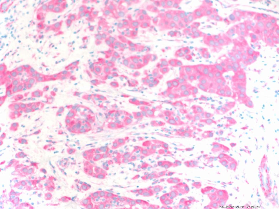
 Photos 200x

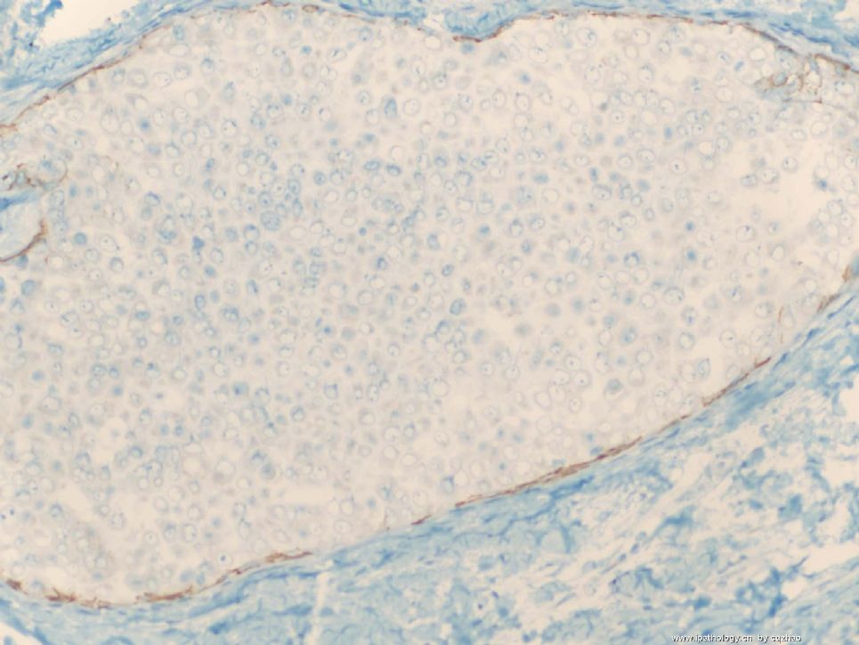
F1 E-cadherine for insitu ca 图1 原位癌E-cadherin染色

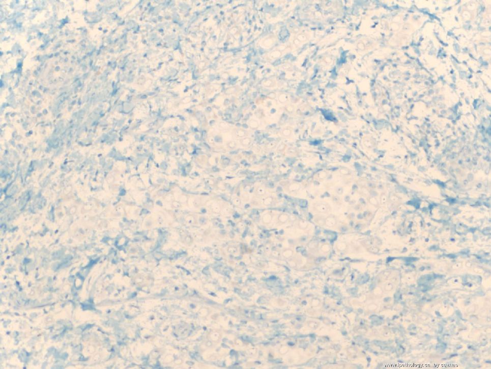
F2 E-cadherine for invasive 图2 浸润癌E-cadherin染色

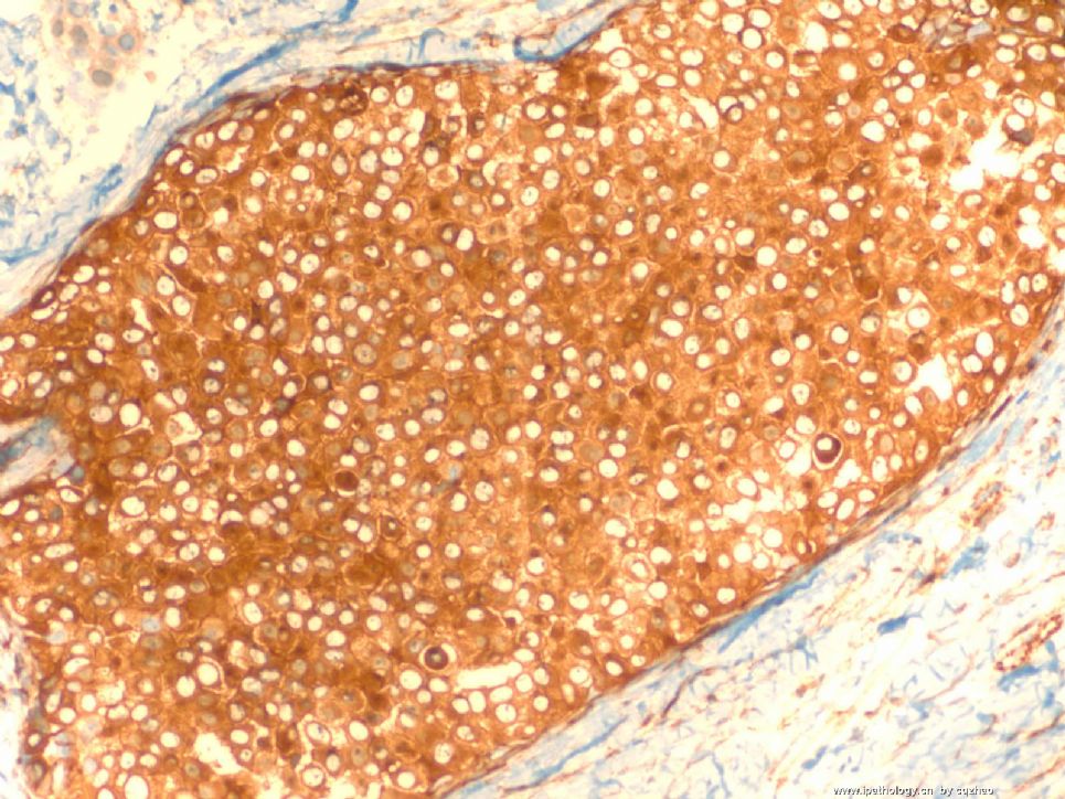
F3 P120 for insitu ca 图3 原位癌P120染色

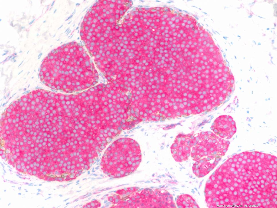
F4. P120 for invasive ca 图4 浸润癌P120染色


Ductal ca (or normal ducts): memberane stain for both e-cad and p120

Lobular lesion: Absence of  stain for E-cad and strong cytoplasmic stain for p120.

导管癌(或正常导管): E-cadherin和p120均呈膜阳性。

小叶病变:E-cadherin不着色,p120胞浆强阳性。

 (abin译)


名称:图1
描述:图1

名称:图2
描述:图2

名称:图3
描述:图3

名称:图4
描述:图4
0
回复

cqzhao 离线

帖子:5518
粉蓝豆:1071
经验:5755
注册时间:2008-09-29
加关注  |  发消息
45 楼    发表于2008-12-14 02:46:00举报|引用
返回顶部 | 快捷回复
本帖最后由 于 2008-12-14 14:06:00 编辑

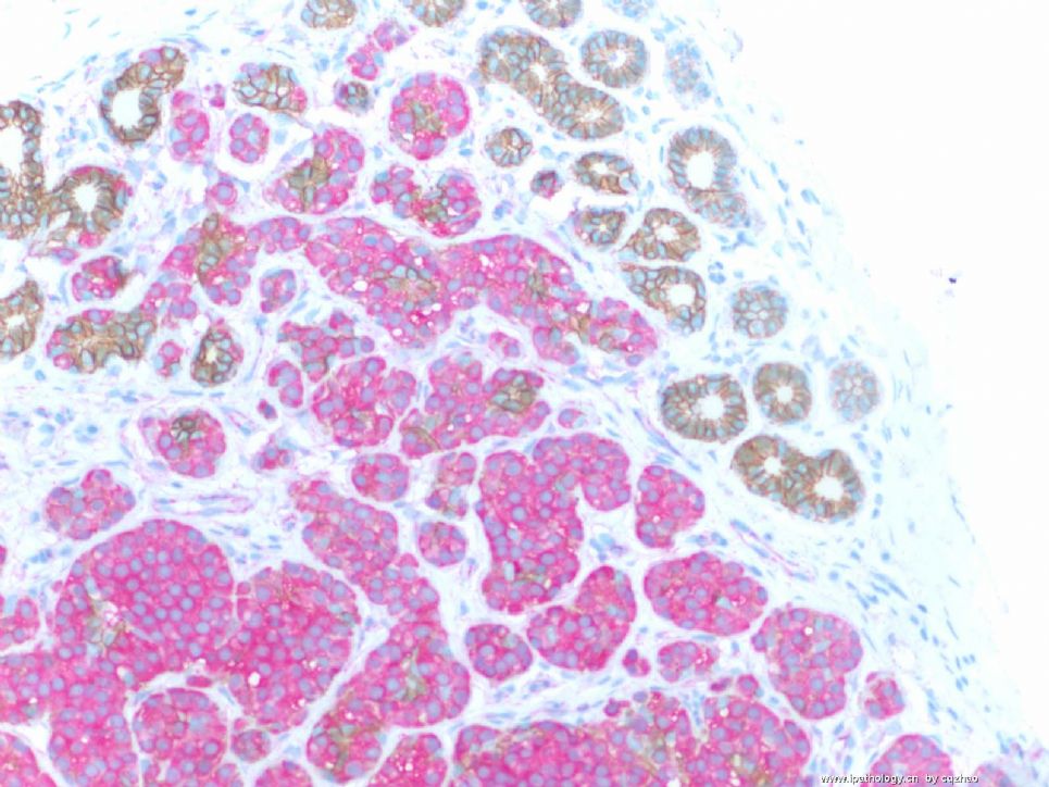
 Dual stains: anti-ecad labeling with brown color, anti-p120 with pink color, mixed stain, one procedure.

F 1 for insi tu ca

F2 for invasive ca

F3 in situ and normal ducts.

E-cad membrane stain (brown)

P-120 cytoplasmic stain (pink)

Lobular lesion will not see brown color and only pick.

Fig 3: some normal ducts show brown membrane stain and no pink color.

So you know the nature of the lesions.

Lobular carcinoma in situ and invasive lobular carcinoma.


Now, what type of invasive lobular carcinoma is it?

abin译:

免疫组化双标记:抗E-cadherin棕色,抗p120粉红色。
图1 原位癌
图2 浸润癌
图3 原位癌和正常导管。E-cadherin呈膜阳性(棕色),P-120呈胞浆阳性(粉红色)
小叶病变不见棕色,只有粉红色。正常导管示棕色的膜染色,没有粉红色。
因此你明白病变的性质了:小叶原位癌和浸润性小叶癌。
那么,是什么亚型的浸润性小叶癌呢?


名称:图1
描述:图1

名称:图2
描述:图2

名称:图3
描述:图3
0
回复

abin 离线

帖子:7383
粉蓝豆:6014
经验:11754
注册时间:2006-10-08
加关注  |  发消息
46 楼    发表于2008-12-14 14:22:00举报|引用
返回顶部 | 快捷回复
以下是引用cqzhao在2008-12-14 0:25:00的发言:

以下是引用天山望月在2008-12-13 23:21:00的发言:

以下是引用cqzhao在2008-12-12 11:50:00的发言:

  I think Abin is right. Most of people here have good English. I guess most people reading in the website are young, 70 after (borned after 1970). Am I right?

Yes,You are right.hehe


People aged 40 above are proffesors or experts already. They do not need to learn.

Maybe they learn by other ways.

0
回复
signature

华夏病理/粉蓝医疗

为基层医院病理科提供全面解决方案,

努力让人人享有便捷准确可靠的病理诊断服务。


abin 离线

帖子:7383
粉蓝豆:6014
经验:11754
注册时间:2006-10-08
加关注  |  发消息
47 楼    发表于2008-12-14 14:49:00举报|引用
返回顶部 | 快捷回复
本帖最后由 于 2008-12-14 14:52:00 编辑

 pleomorphic variant?

Some invasive lobular carcinomas consist entirely or in part of cells larger than cells in classical invasive lobular carcinoma with relatively abundant, eosinophilic cytoplasm (Fig. 32.17). The nucleus in some examples is hyperchromatic and eccentric with a distinct nucleolus creating a plasmacytoid appearance (Fig. 32.18). These cells have been referred to variously as myoid (12), histiocytoid (77-79), and pleomorphic lobular carcinoma (80,81) (Figs. 32.18-19). __from Rosen's Breat Pathology (P700)

0
回复
signature

华夏病理/粉蓝医疗

为基层医院病理科提供全面解决方案,

努力让人人享有便捷准确可靠的病理诊断服务。


海马 离线

帖子:504
粉蓝豆:12
经验:556
注册时间:2008-08-26
加关注  |  发消息
48 楼    发表于2008-12-14 16:26:00举报|引用
返回顶部 | 快捷回复
 浸润性导管癌
0
回复

cqzhao 离线

帖子:5518
粉蓝豆:1071
经验:5755
注册时间:2008-09-29
加关注  |  发消息
49 楼    发表于2008-12-14 22:12:00举报|引用
返回顶部 | 快捷回复
本帖最后由 于 2008-12-16 19:01:00 编辑
以下是引用海马在2008-12-14 16:26:00的发言:

 浸润性导管癌

friend, please check the IHC photos and my interpretation of the IHC result.

abin译:

朋友,请核对免疫组化照片和我对免疫组化结果的解释(23楼)

0
回复

天山望月 离线

帖子:4902
粉蓝豆:83
经验:5092
注册时间:2007-02-03
加关注  |  发消息
50 楼    发表于2008-12-14 23:35:00举报|引用
返回顶部 | 快捷回复

 Wow............ ,So beautiful the photos!Thanks !

It is time to go, I will answer the question tomorrow.

0
回复
signature
广州金域病理

天山望月 离线

帖子:4902
粉蓝豆:83
经验:5092
注册时间:2007-02-03
加关注  |  发消息
51 楼    发表于2008-12-15 19:05:00举报|引用
返回顶部 | 快捷回复
以下是引用cqzhao在2008-12-14 2:46:00的发言:


Now, what type of invasive lobular carcinoma is it?

那么,是什么亚型的浸润性小叶癌呢?

先复习浸润性小叶癌的分型,再回答问题。

先描述一下浸润性小叶癌(ILC)的细胞形态特点:

A、经典型的细胞特点:多为黏附性差(即松散)的圆形卵圆形小细胞,浆少,核偏位,圆形,核仁不明显,核分裂少,可见胞浆内小管腔。

B、多形性小叶癌细胞:浆细胞样、黏液印戒样、组织细胞样或大汗腺样分化等。

ILC分为:

1、经典型:具有ILC经典型特征的瘤细胞,成列兵样、靶环状、单个散在浸润在间质纤维组织中,>90%伴LCIS.

2、变型,WHO 在变型中提出四个亚型:

1)、腺泡型至少20个以上的具有ILC经典型特征的瘤细胞成团状、簇状聚集,浸润间质。

2)、实性型:具有ILC经典型特征的瘤细胞弥漫成片浸润间质,多形性较经典型明显,核分裂较多,间质纤维组织少。

3)、多形型:保持小叶癌的生长方式,非典型性和多形性更明显。常出现多形性小叶癌细胞。

4)、混合型:有经典型和一种或一种以上的亚型复合组成。

只有80%以上区域表现某一形态特点的病例才能归为某一特殊类型。

0
回复
signature
广州金域病理

天山望月 离线

帖子:4902
粉蓝豆:83
经验:5092
注册时间:2007-02-03
加关注  |  发消息
52 楼    发表于2008-12-15 19:18:00举报|引用
返回顶部 | 快捷回复

 此例有经典型(散在小细胞)、实性型(图2、3,密集小细胞区),、比经典型细胞大的细胞区(图6、7组织细胞样分化,多形性明显)。

考虑:多形性或混合型,倾向混合型可能性大。

不知当否,请专家赐教!谢谢!

0
回复
signature
广州金域病理

student 离线

帖子:16
粉蓝豆:1
经验:27
注册时间:2008-06-30
加关注  |  发消息
53 楼    发表于2008-12-16 19:09:00举报|引用
返回顶部 | 快捷回复
以下是引用中原老狼在2008-12-15 19:41:00的发言:

 ca

老师讲解这么多了,这两个字母令人失望。

网上讨论不管对错,不分水平高下。但是,对于教学病例的讨论,学习并思考后再发言是对老师的尊重。

0
回复

stevenshen 离线

帖子:343
粉蓝豆:2
经验:343
注册时间:2008-06-03
加关注  |  发消息
54 楼    发表于2008-12-06 09:02:00举报|引用
返回顶部 | 快捷回复
本帖最后由 于 2008-12-10 20:00:00 编辑

It is possible that it is invasive ductal carcinoma and solid DCIS; I would guess that it is LCIS with invasive lobular carcinoma (pleomorphic type).  Thanks.  Looking forward to hearing the discussion and final answer.

(abin译:这例可能是浸润性导管癌和实性DCIS。我猜也可能是LCIS伴浸润性小叶癌(多形性亚型)。谢谢。期待听到讨论和最终结果。)

0
回复

cqzhao 离线

帖子:5518
粉蓝豆:1071
经验:5755
注册时间:2008-09-29
加关注  |  发消息
55 楼    发表于2008-12-09 11:58:00举报|引用
返回顶部 | 快捷回复
本帖最后由 于 2008-12-10 20:01:00 编辑
以下是引用杨宝军在2008-12-3 21:00:00的发言:

 是否为穿刺活检组织,局部好像有菊形团结构,首选浸润性导管癌伴有神经内分泌分化


It is core biopsy specimen and  a consult case from other hospital.

(abin译:这是粗针穿刺活检标本,是来自其他医院的会诊病例。)

0
回复

abin 离线

帖子:7383
粉蓝豆:6014
经验:11754
注册时间:2006-10-08
加关注  |  发消息
56 楼    发表于2008-12-10 20:03:00举报|引用
返回顶部 | 快捷回复
 诚如Dr.Stevenshen所述,考虑原位癌伴浸润癌。是DCIS伴IDC还是LCIS伴ILC,需要先做免疫组化:E-Ca和p120
0
回复
signature

华夏病理/粉蓝医疗

为基层医院病理科提供全面解决方案,

努力让人人享有便捷准确可靠的病理诊断服务。


cqzhao 离线

帖子:5518
粉蓝豆:1071
经验:5755
注册时间:2008-09-29
加关注  |  发消息
57 楼    发表于2008-12-11 02:00:00举报|引用
返回顶部 | 快捷回复

 Good analysis. H&E slides are good enough to know the presence of insitu and invasive carcinoma. The key for this case is the nature of the tumor. I will take some photos when I have time.

You can continue to guess.

0
回复

qxysjsh 离线

帖子:73
粉蓝豆:19
经验:85
注册时间:2008-08-07
加关注  |  发消息
58 楼    发表于2008-12-11 12:03:00举报|引用
返回顶部 | 快捷回复
 浸润性导管癌
0
回复

闲来看云 离线

帖子:179
粉蓝豆:42
经验:2005
注册时间:2006-09-27
加关注  |  发消息
59 楼    发表于2008-12-11 16:33:00举报|引用
返回顶部 | 快捷回复
以下是引用cqzhao在2008-12-11 2:00:00的发言:

 Good analysis. H&E slides are good enough to know the  of insitu and invasive carcinoma. The key for this case is the nature of the tumor. I will take some photos when I have time.

You can continue to guess.

分析的好,HE切片足以识别原位癌和浸润癌,这个病例的关键是肿瘤的特性,有时间时,我会采一些图片,

大家继续讨论

闲来看云译

0
回复

wy1992 离线

帖子:4855
粉蓝豆:1
经验:6825
注册时间:2007-06-30
加关注  |  发消息
60 楼    发表于2008-12-12 12:12:00举报|引用
返回顶部 | 快捷回复
 I AM BEFORE 1970.AND i THINK THIS CASE SEEMS TO BE CARCINOMA IN SITU WITHOUT DEFININTE INVASIVENESS
0
回复
signature

朱正龙

回复:105 阅读:10127
共6页/105条首页上一页12345下一页尾页
【免责声明】讨论内容仅作学术交流之用,不作为诊疗依据,由此而引起的法律问题作者及本站不承担任何责任。
快速回复
进入高级回复
您最多可输入10000个汉字,按 "Ctrl" + "Enter" 直接发送
搜索回复/乘电梯 ×
按内容
按会员
乘电梯
合作伙伴
友情链接