| 图片: | |
|---|---|
| 名称: | |
| 描述: | |
- Breast lobular lesions and stains ( cqz 7)
-
本帖最后由 于 2008-12-23 23:04:00 编辑
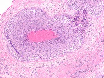
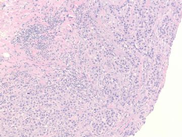
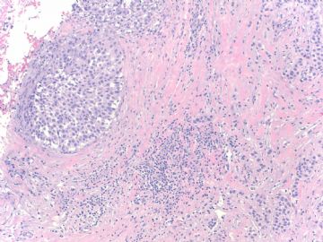
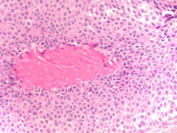
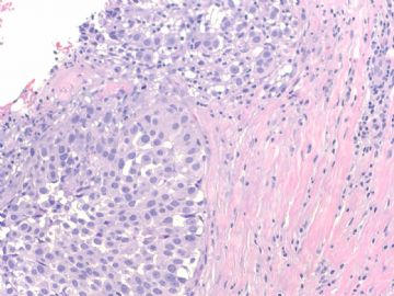
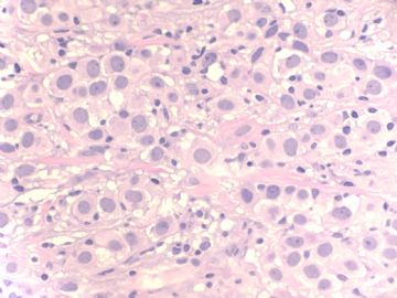
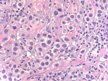
young women (<30 y) had breast reduction. Two sections for each sides were submitted for microscopic examination. See photo for one section. How will you sign out the case?
abin译:年轻女性(<30岁),乳房缩小手术标本。一张切片的两个部位的显微镜下检查。先看第一个部位。你会怎样签发报告?
Fig
200x
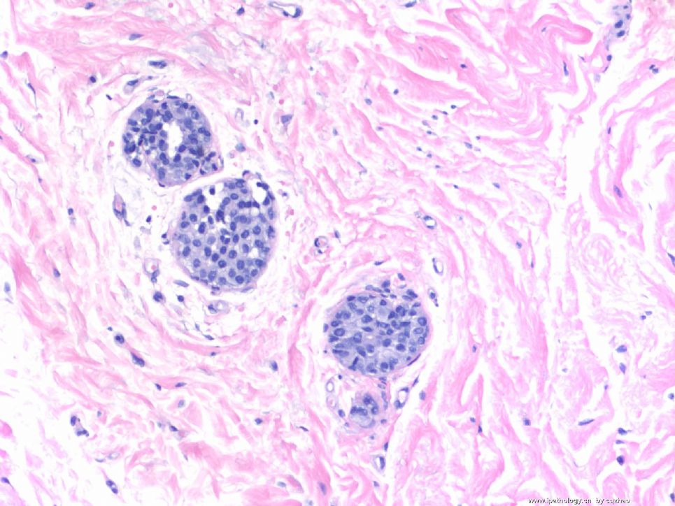
名称:图1
描述:图1
| 以下是引用天山望月在2008-12-13 23:21:00的发言:
|
People aged 40 above are proffesors or experts already. They do not need to learn.
-
本帖最后由 于 2008-12-14 14:06:00 编辑
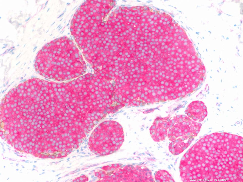
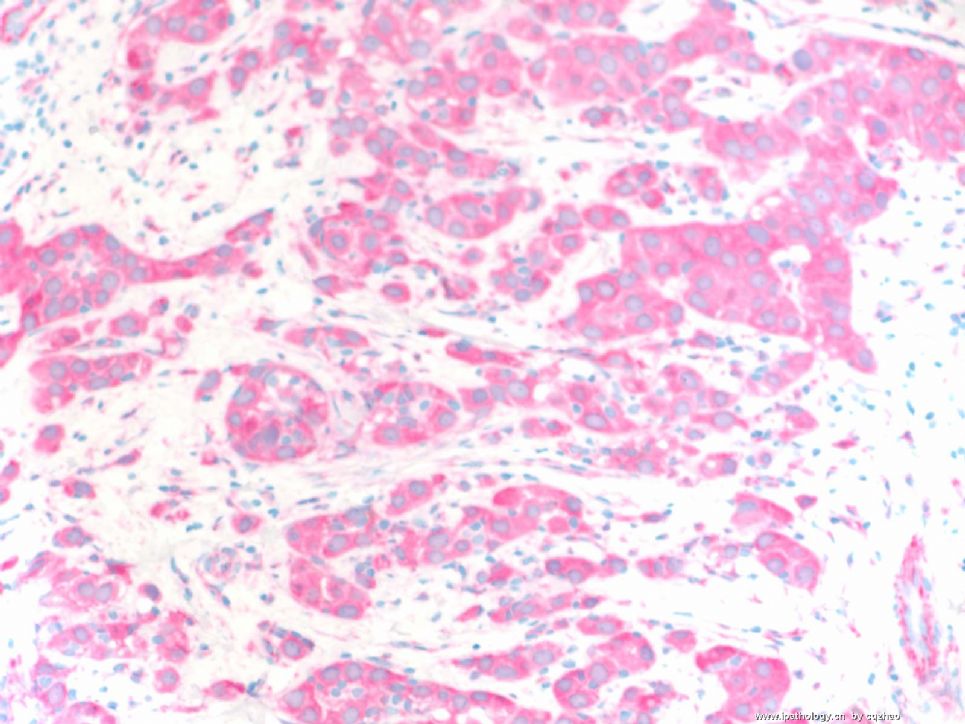
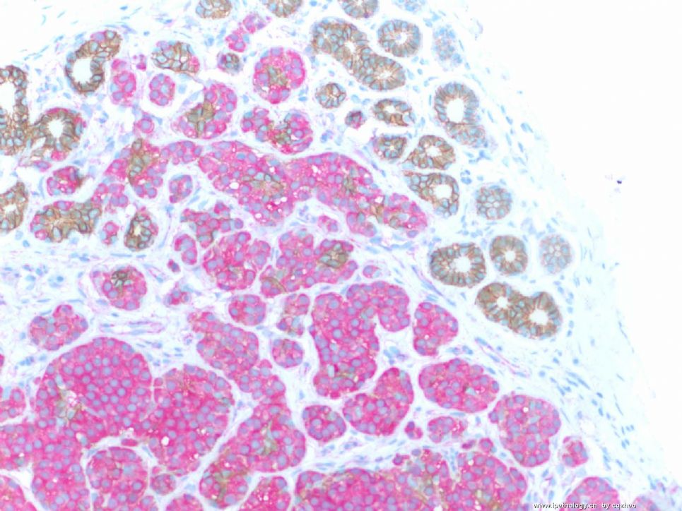
Dual stains: anti-ecad labeling with brown color, anti-p120 with pink color, mixed stain, one procedure.
F 1 for insi tu ca
F2 for invasive ca
F3 in situ and normal ducts.
E-cad membrane stain (brown)
P-120 cytoplasmic stain (pink)
Lobular lesion will not see brown color and only pick.
Fig 3: some normal ducts show brown membrane stain and no pink color.
So you know the nature of the lesions.
Lobular carcinoma in situ and invasive lobular carcinoma.
Now, what type of invasive lobular carcinoma is it?
abin译:
免疫组化双标记:抗E-cadherin棕色,抗p120粉红色。
图1 原位癌
图2 浸润癌
图3 原位癌和正常导管。E-cadherin呈膜阳性(棕色),P-120呈胞浆阳性(粉红色)
小叶病变不见棕色,只有粉红色。正常导管示棕色的膜染色,没有粉红色。
因此你明白病变的性质了:小叶原位癌和浸润性小叶癌。
那么,是什么亚型的浸润性小叶癌呢?
名称:图1
描述:图1
名称:图2
描述:图2
名称:图3
描述:图3
-
本帖最后由 于 2008-12-14 14:51:00 编辑
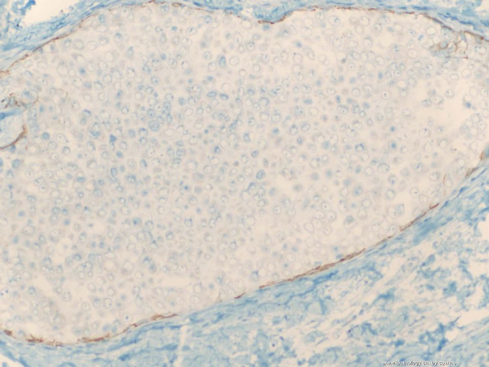
Photos 200x
F1 E-cadherine for insitu ca 图1 原位癌E-cadherin染色
F2 E-cadherine for invasive 图2 浸润癌E-cadherin染色
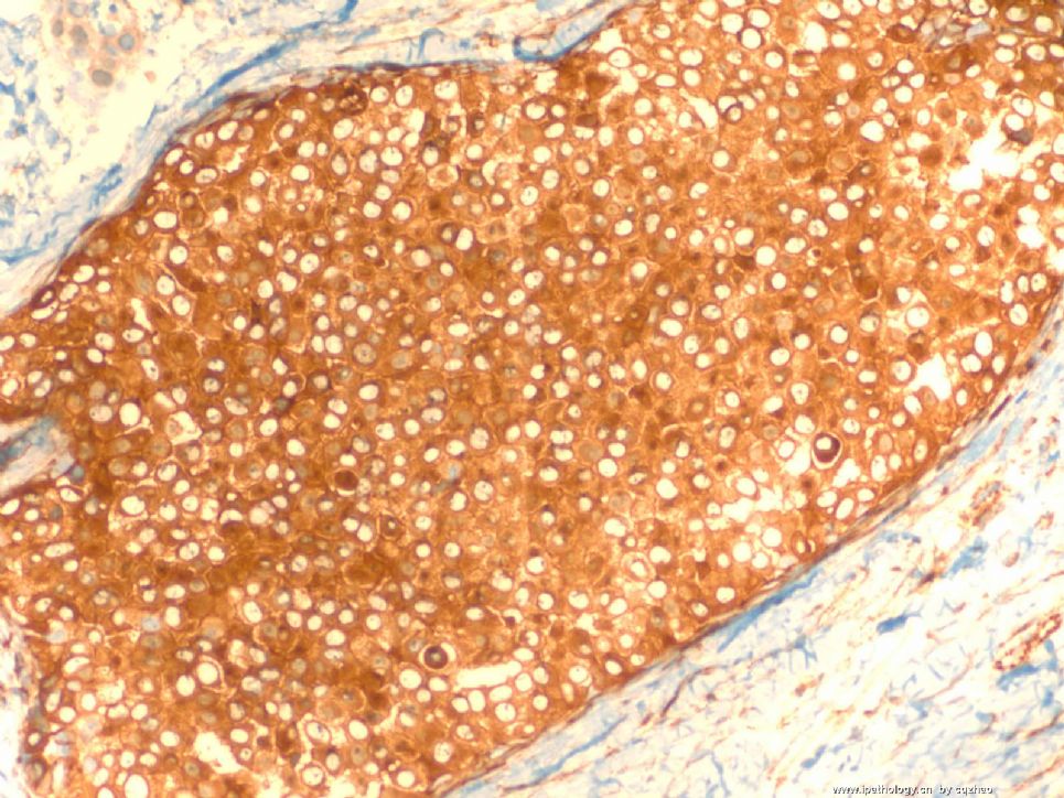
F3 P120 for insitu ca 图3 原位癌P120染色
F4. P120 for invasive ca 图4 浸润癌P120染色
Ductal ca (or normal ducts): memberane stain for both e-cad and p120
Lobular lesion: Absence of stain for E-cad and strong cytoplasmic stain for p120.
导管癌(或正常导管): E-cadherin和p120均呈膜阳性。
小叶病变:E-cadherin不着色,p120胞浆强阳性。
(abin译)
名称:图1
描述:图1

名称:图2
描述:图2
名称:图3
描述:图3

名称:图4
描述:图4
