共2页/30条首页上一页12下一页尾页
回复:30 阅读:9803
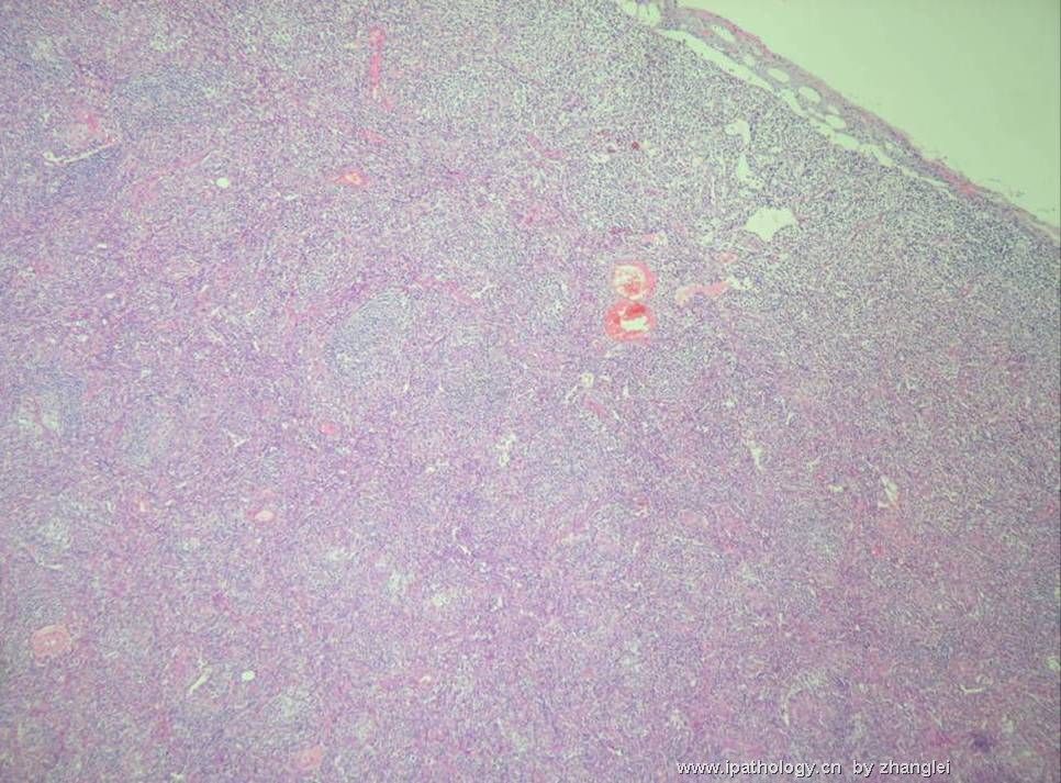
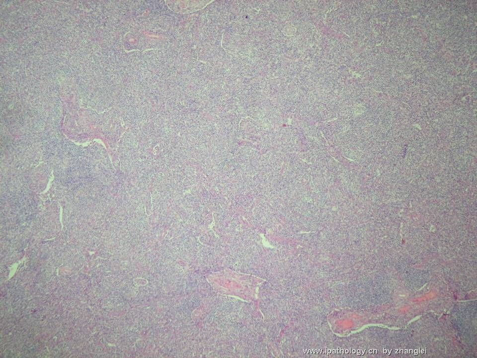
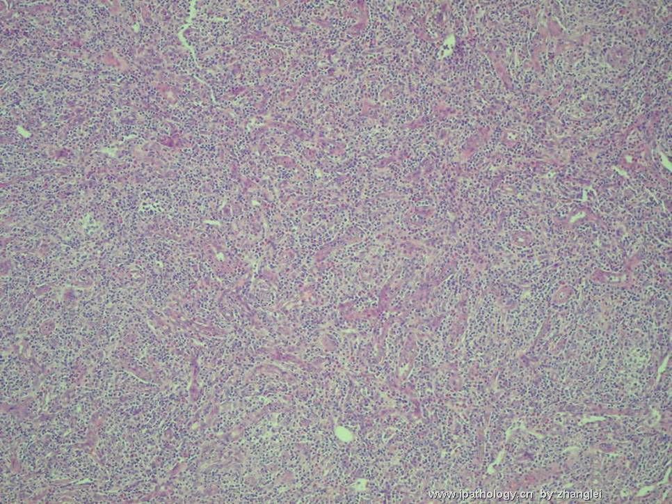
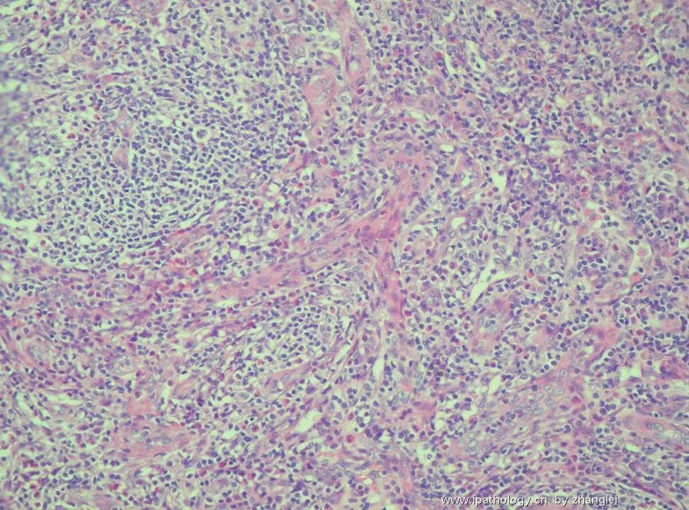
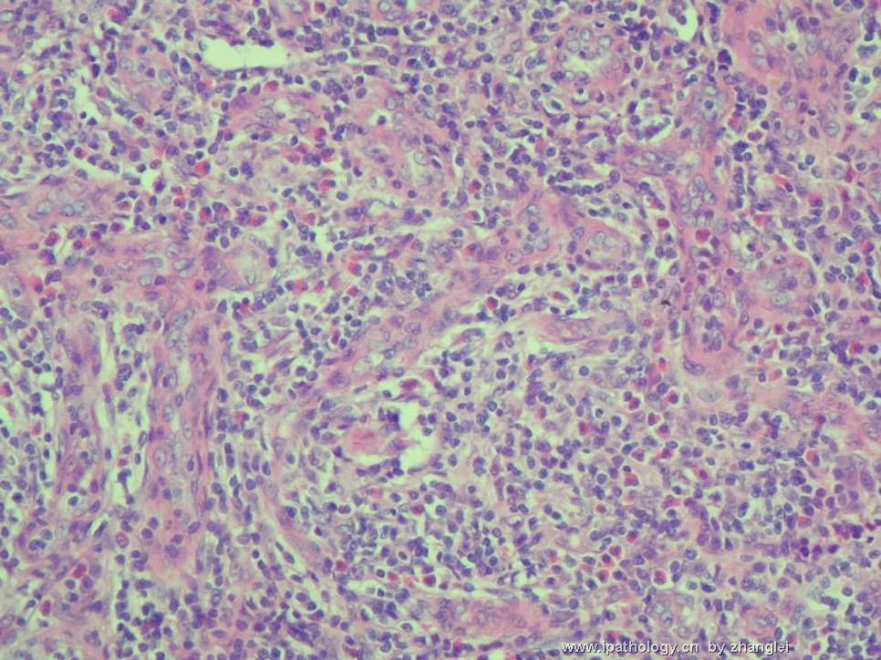
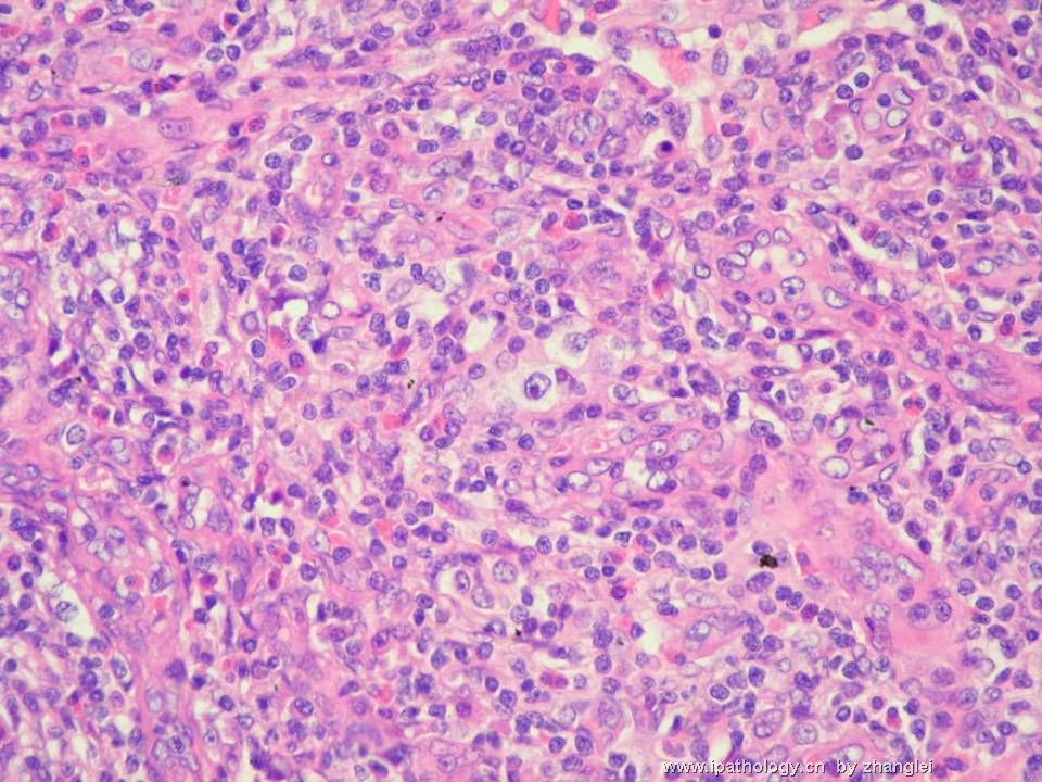
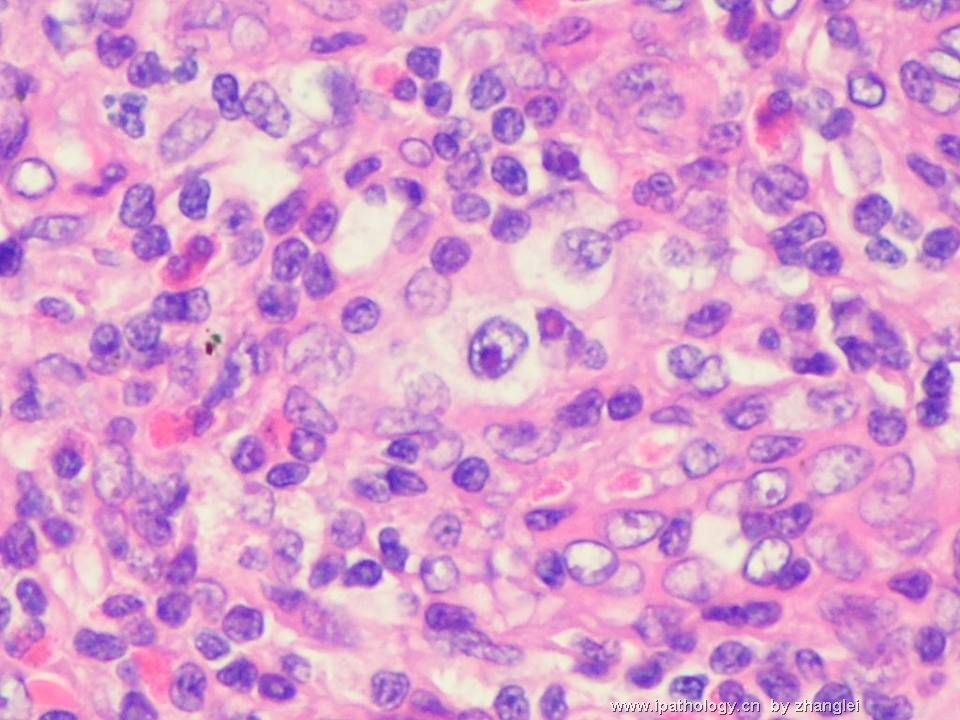
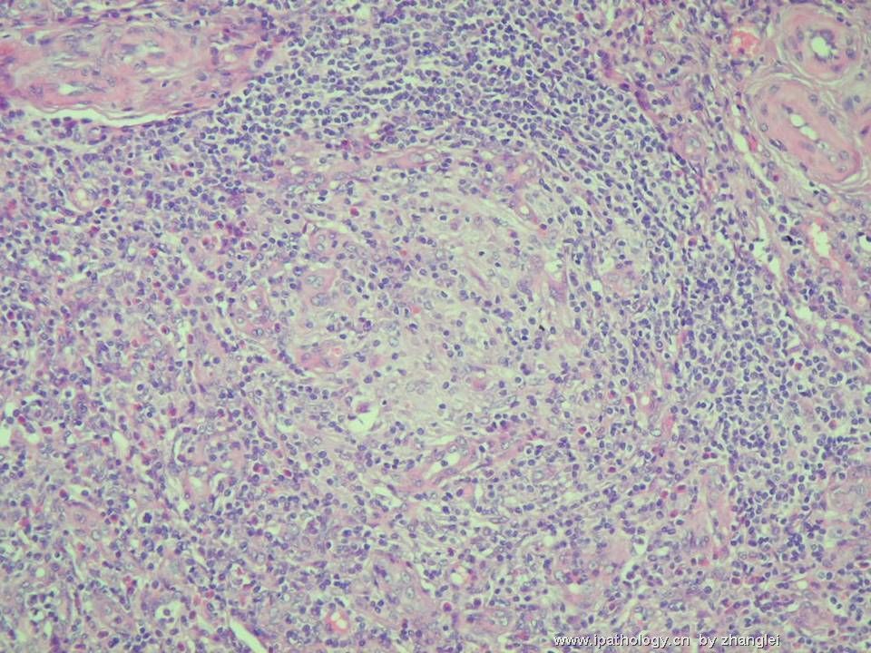
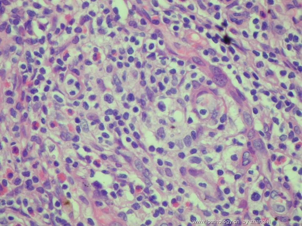
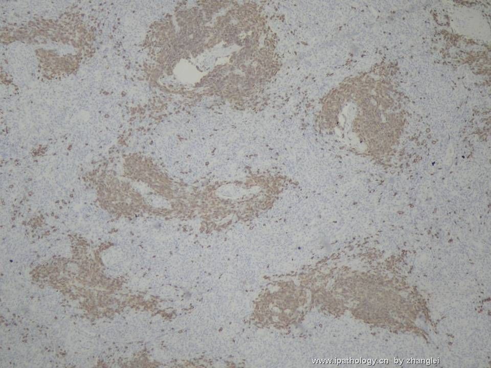
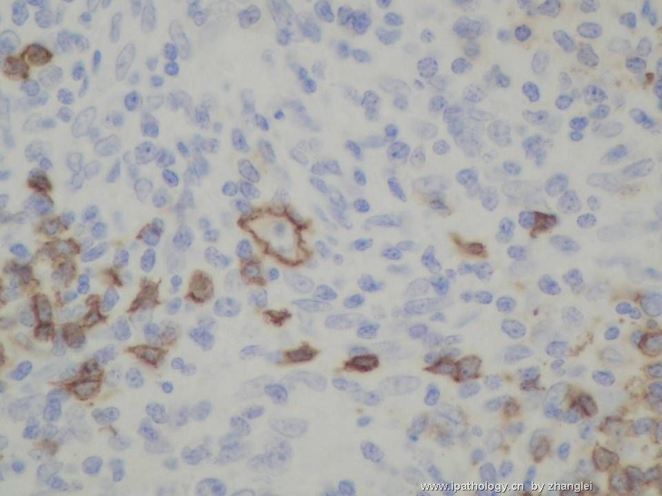
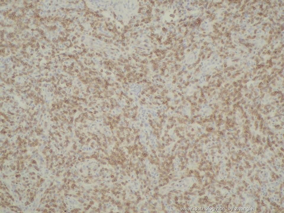
(转贴)颈部淋巴结--血管免疫母细胞性T细胞淋巴瘤(angioimmunoblastic T cell lymphoma)

zhanglei 离线

帖子:3751
粉蓝豆:2078
经验:3901
注册时间:2006-09-26
加关注  |  发消息
楼主 发表于 2007-03-04 17:56|举报|关注(4)
浏览排序[ 顺序 逆序 楼主 支持 精彩 ]  快捷回复
姓    名: ××× 性别:   年龄:  53
标本名称:  颈部淋巴结
简要病史:  间断高烧40余天,住院发现全身淋巴结肿大。
肉眼检查:  
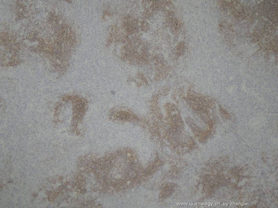
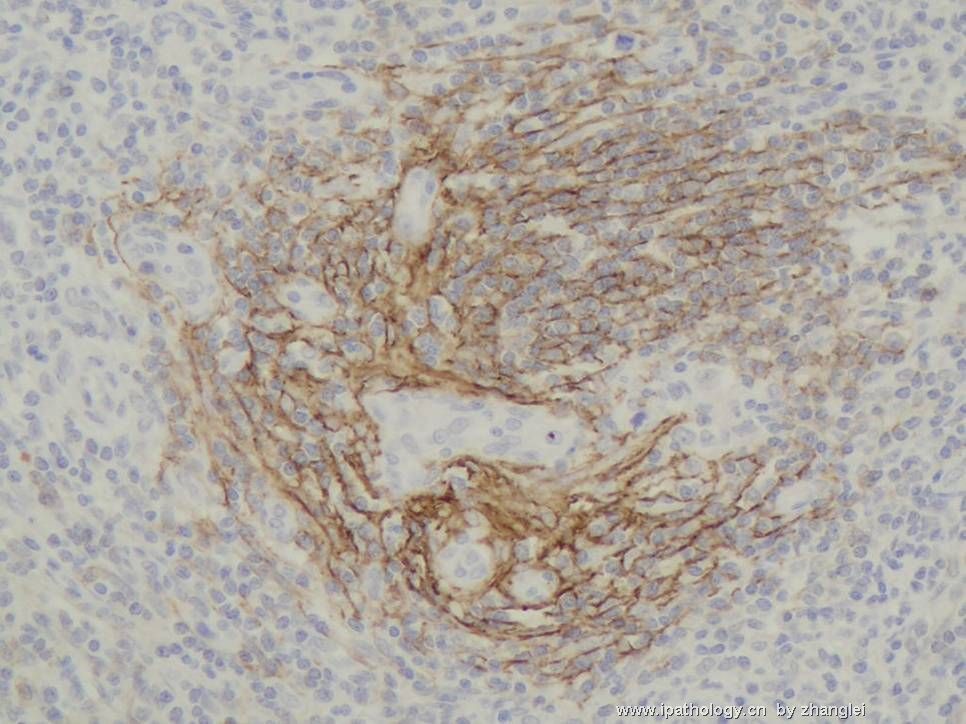
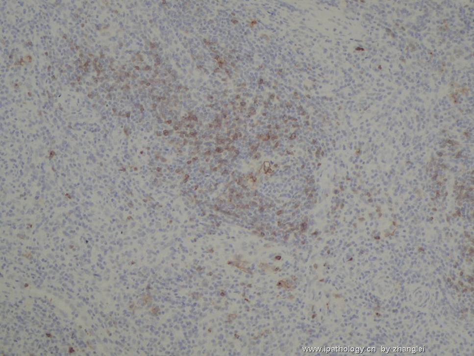
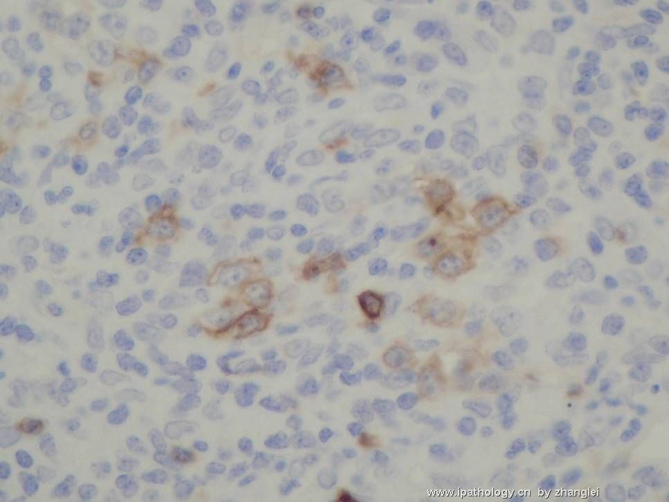
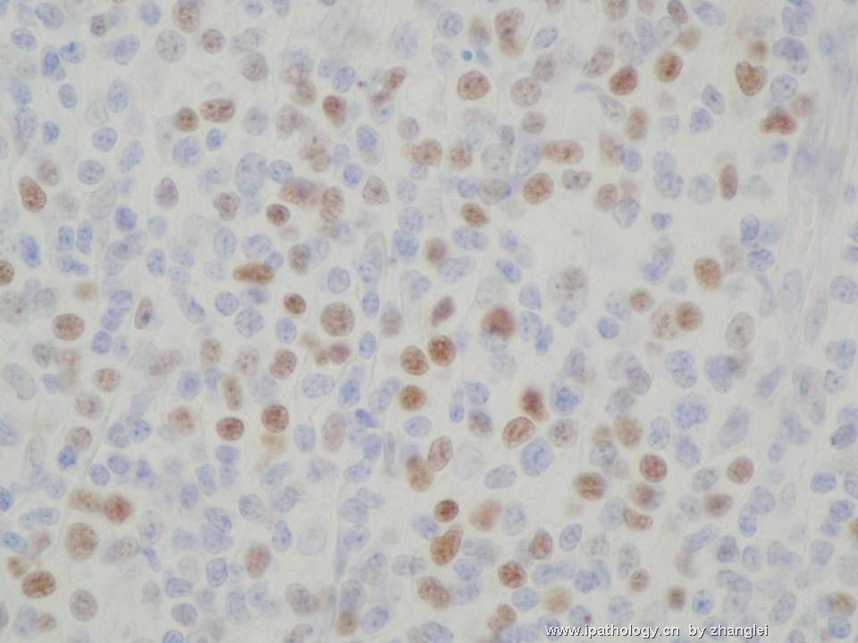
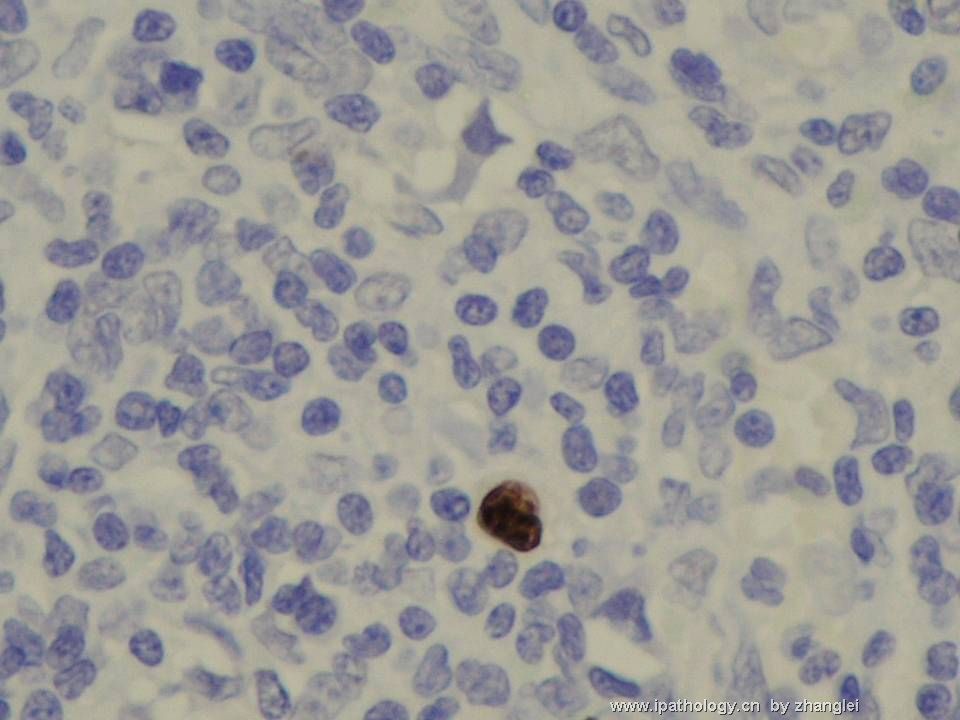
图11、12--CD20,13--CD3,14、15—CD21,16、17—CD10,18――BCL-6,19---EBER
(转贴)颈部淋巴结--血管免疫母细胞性T细胞淋巴瘤(angioimmunoblastic T cell lymphoma)图1
名称:图1
描述:图1
(转贴)颈部淋巴结--血管免疫母细胞性T细胞淋巴瘤(angioimmunoblastic T cell lymphoma)图2
名称:图2
描述:图2
(转贴)颈部淋巴结--血管免疫母细胞性T细胞淋巴瘤(angioimmunoblastic T cell lymphoma)图3
名称:图3
描述:图3
(转贴)颈部淋巴结--血管免疫母细胞性T细胞淋巴瘤(angioimmunoblastic T cell lymphoma)图4
名称:图4
描述:图4
(转贴)颈部淋巴结--血管免疫母细胞性T细胞淋巴瘤(angioimmunoblastic T cell lymphoma)图5
名称:图5
描述:图5
(转贴)颈部淋巴结--血管免疫母细胞性T细胞淋巴瘤(angioimmunoblastic T cell lymphoma)图6
名称:图6
描述:图6
(转贴)颈部淋巴结--血管免疫母细胞性T细胞淋巴瘤(angioimmunoblastic T cell lymphoma)图7
名称:图7
描述:图7
(转贴)颈部淋巴结--血管免疫母细胞性T细胞淋巴瘤(angioimmunoblastic T cell lymphoma)图8
名称:图8
描述:图8
(转贴)颈部淋巴结--血管免疫母细胞性T细胞淋巴瘤(angioimmunoblastic T cell lymphoma)图9
名称:图9
描述:图9
(转贴)颈部淋巴结--血管免疫母细胞性T细胞淋巴瘤(angioimmunoblastic T cell lymphoma)图10
名称:图10
描述:图10
(转贴)颈部淋巴结--血管免疫母细胞性T细胞淋巴瘤(angioimmunoblastic T cell lymphoma)图11
名称:图11
描述:图11
(转贴)颈部淋巴结--血管免疫母细胞性T细胞淋巴瘤(angioimmunoblastic T cell lymphoma)图12
名称:图12
描述:图12
(转贴)颈部淋巴结--血管免疫母细胞性T细胞淋巴瘤(angioimmunoblastic T cell lymphoma)图13
名称:图13
描述:图13
(转贴)颈部淋巴结--血管免疫母细胞性T细胞淋巴瘤(angioimmunoblastic T cell lymphoma)图14
名称:图14
描述:图14
(转贴)颈部淋巴结--血管免疫母细胞性T细胞淋巴瘤(angioimmunoblastic T cell lymphoma)图15
名称:图15
描述:图15
(转贴)颈部淋巴结--血管免疫母细胞性T细胞淋巴瘤(angioimmunoblastic T cell lymphoma)图16
名称:图16
描述:图16
(转贴)颈部淋巴结--血管免疫母细胞性T细胞淋巴瘤(angioimmunoblastic T cell lymphoma)图17
名称:图17
描述:图17
(转贴)颈部淋巴结--血管免疫母细胞性T细胞淋巴瘤(angioimmunoblastic T cell lymphoma)图18
名称:图18
描述:图18
(转贴)颈部淋巴结--血管免疫母细胞性T细胞淋巴瘤(angioimmunoblastic T cell lymphoma)图19
名称:图19
描述:图19
标签:
本帖最后由 于 2007-03-13 22:08:00 编辑
0
×参考诊断
血管免疫母细胞性T细胞淋巴瘤

liuyong 离线

帖子:213
粉蓝豆:517
经验:218
注册时间:2006-09-20
加关注  |  发消息
21 楼    发表于2007-03-05 01:32:00举报|引用
返回顶部 | 快捷回复
以下是引用zhongshihua 在2007-3-4 20:58:00的发言:

 考虑血管免疫母细胞性T细胞淋巴瘤。


血管免疫母细胞性T细胞淋巴瘤(angioimmunoblastic T cell lymphoma)
1、临床表现:老年人,男性多见,全身淋巴结肿大,发热。
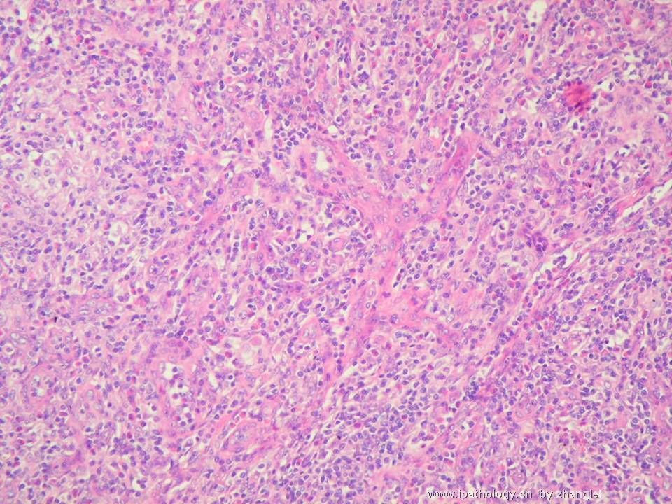
2、镜下形态:淋巴结结构基本破坏,淋巴滤泡萎缩减少,分枝状小血管增生明显,血管基膜增厚,形态大小不一致的淋巴样细胞弥漫增生,其中见散在的免疫母细胞,胞质丰富淡染,胞核圆形或稍不规则形,核膜厚,核仁明显。可见少量散在的嗜酸性粒细胞。
3、免疫表型:CD20- 、CD3+ 、CD21显示血管旁的FDC网、CD10+ 、BCL-6+ 、EBER大细胞+
临床表现、镜下形态和免疫表型均符合诊断血管免疫母细胞性T细胞淋巴瘤(angioimmunoblastic T cell lymphoma)
0
回复

abin 离线

帖子:7416
粉蓝豆:6014
经验:11787
注册时间:2006-10-08
加关注  |  发消息
22 楼    发表于2007-03-05 20:26:00举报|引用
返回顶部 | 快捷回复
 谢谢楼主和楼上各位老师!特别感谢刘老师讲解!
0
回复
signature

华夏病理/粉蓝医疗

为基层医院病理科提供全面解决方案,

努力让人人享有便捷准确可靠的病理诊断服务。


国泰民安 离线

帖子:231
粉蓝豆:8
经验:743
注册时间:2006-11-01
加关注  |  发消息
23 楼    发表于2007-04-04 21:52:00举报|引用
返回顶部 | 快捷回复
 典型病例
0
回复

xiaohl 离线

帖子:282
粉蓝豆:24
经验:288
注册时间:2006-10-19
加关注  |  发消息
24 楼    发表于2007-04-05 13:37:00举报|引用
返回顶部 | 快捷回复
 好典型
0
回复

emmagao 离线

帖子:169
粉蓝豆:42
经验:447
注册时间:2007-12-16
加关注  |  发消息
25 楼    发表于2008-08-02 16:41:00举报|引用
返回顶部 | 快捷回复
 THANKS FOR SHARING!
0
回复

naonao 离线

帖子:35
粉蓝豆:1931
经验:70
注册时间:2008-07-08
加关注  |  发消息
26 楼    发表于2008-07-08 07:12:00举报|引用
返回顶部 | 快捷回复
 好,相见恨晚
0
回复
signature

panzenggang 离线

帖子:189
粉蓝豆:480
经验:246
注册时间:2008-01-09
加关注  |  发消息
27 楼    发表于2008-07-08 10:38:00举报|引用
返回顶部 | 快捷回复
 

Angioimmunoblastic T-cell lymphoma

The Key Features

  • Diffuse paracortical infiltrate of polymorphous neoplastic T cells

  • Prominent proliferation of high endothelial venules

  • Proliferation of follicular dendritic cells

CLINICAL FEATURES

  • A peripheral T-cell lymphoma with a polymorphous infiltrate in lymph node, a prominent proliferation of high endothelial venules and follicular dendritic cells;

  • Age: middle to elderly; Gender: M=F;

  • 15-20% of peripheral T-cell lymphoma, 1-2% of non-Hodgkin lymphoma;

  • Clinical presentations: often generalized peripheral lymphadenopathy, hepatosplenomegaly, frequent skin rash, and commonly bone marrow involvement upon biopsy.

MICROSCOPIC FINDINGS

  • Loss of normal lymph node architecture
    • Diffuse paracortical infiltrate of polymorphous neoplastic T cells
    • Lymph node architecture is partially or totally effaced
    • Lymphoid follicles, hyperplastic, depleted or regressed, with irregular borders and lack of mantle zones
  • Neoplastic T cells
    • Small to medium-sized but occasionally large cells, usually show minimal cytologic atypia
    • Abundant clear to pale cytoplasm, distinct cell membranes and irregular nuclear contour
    • Often in clusters around high endothelial venules
    • Often obscured by reactive lymphocytes, immunoblasts, plasma cells, histiocytes, and eosinophils
  • Prominent proliferation high endothelial venules
    • Prominent arborizing high endothelial venules with PAS positive amorphous perivascular material
    • The nuclei of endothelial cells are round to oval with regular nuclear contour and a small central nucleolus
  • Prominent proliferation of follicular dendritic cells
    • Follicular dendritic cell proliferation outside germinal centers/around high endothelial venules
    • Highlighted by CD21 staining
  • B cell proliferation
    • >70% are EBV positive
    • May be polymorphic or monomorphic, immunoblastic or plasmacytic
    • Immunoglobulin gene rearrangement detected in 10% cases
    • May produce a Hodgkin-like proliferation with Reed-Sternberg-like cells

DIFFERENTIAL DIAGNOSES

  • Reactive lymphadenopathies

  • Multicentric Castleman's disease

  • Diffuse large B-cell lymphoma

  • Classical Hodgkin's Disease

IMMUNOHISTOCHEMISTRY AND SPECIAL STAINS

  • Neoplastic cells are mature T-cells with CD3+, CD4+

  • In most cases, the neoplastic T-cells show aberrant expression of CD10

  • Loss of T-cell antigen such as CD7 can occur in some cases

  • EBV positive in >75% cases, mostly in B-cells, not T-cells

  • Follicular dendritic cells CD21+

CYTOGENETICS
  • •TCR gene rearrangement in 75% cases
  • 90% have cytogenetic alterations: trisomy 3, trisomy 5 and gain of chromosome X

TREATMENT AND PROGNOSIS

  • Aggressive, median survival <3 years

REFERENCES

  • Jaffe ES, Harris NL, Stein H, Vardiman JW, editors. Pathology and genetics of tumours of haematopoietic and lymphoid tissues. World Health Organization classification of tumours. Lyon (France): IARC Press; 2001.

  • Practical Diagnosis of Hematologic Disorders, Fourth Edition. By Carl R. Kjeldsberg, 2006.
Summarized by Zenggang Pan, MD, PhD
0
回复

fuying 离线

帖子:362
粉蓝豆:2
经验:390
注册时间:2008-04-10
加关注  |  发消息
28 楼    发表于2008-07-08 11:30:00举报|引用
返回顶部 | 快捷回复
 典型病例,谢谢提供,学习了。
0
回复

nanfeiyan 离线

帖子:152
粉蓝豆:26
经验:235
注册时间:2006-12-10
加关注  |  发消息
29 楼    发表于2007-05-14 21:30:00举报|引用
返回顶部 | 快捷回复
 嗯!有所收获。
0
回复
signature
病理人

stgowest 离线

帖子:237
粉蓝豆:2345
经验:1134
注册时间:2008-03-21
加关注  |  发消息
30 楼    发表于2008-08-06 13:35:00举报|引用
返回顶部 | 快捷回复
 好病立,好讲解。好老师。
0
回复
回复:30 阅读:9803
共2页/30条首页上一页12下一页尾页
【免责声明】讨论内容仅作学术交流之用,不作为诊疗依据,由此而引起的法律问题作者及本站不承担任何责任。
快速回复
进入高级回复
您最多可输入10000个汉字,按 "Ctrl" + "Enter" 直接发送
搜索回复/乘电梯 ×
按内容
按会员
乘电梯
合作伙伴
友情链接