| 图片: | |
|---|---|
| 名称: | |
| 描述: | |
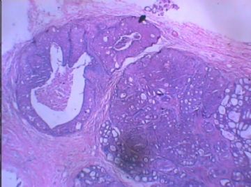
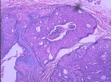
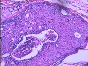
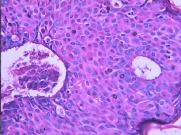
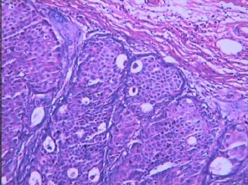
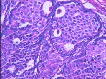
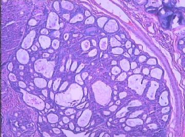
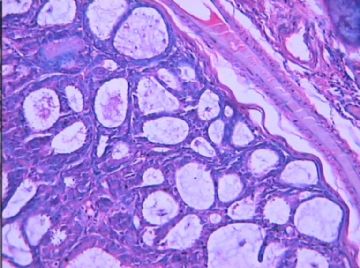
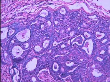
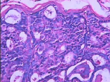
- B1572乳腺低级别导管内癌?
| 姓 名: | ××× | 性别: | 女 | 年龄: | 36岁 |
| 标本名称: | 左侧乳腺包块 | ||||
| 简要病史: | |||||
| 肉眼检查: | |||||
相关帖子
- • 左乳腺肿物
- • 乳腺癌?
- • 乳腺肿物
- • 乳腺肿物
- • 左乳癌标本乳头一个导管内的病变
- • 乳腺两个相邻导管内的病变
- • 乳腺肿物,请各位老师帮忙会诊
- • 女 46岁发现左乳腺肿块一月余
- • 乳腺肿物
- • 乳腺肿物
-
stevenshen 离线
- 帖子:343
- 粉蓝豆:2
- 经验:343
- 注册时间:2008-06-03
- 加关注 | 发消息
I am not confident this is clearly DCIS; I favor florid UDH with adenosis and apocrine changes.
It is true that comedo type necrosis is often associated with high grade DCIS.
I have seen example of florid UDH with small focus central "necrosis".
The last 4 photos are not DCIS; the slides also have drying artifact.
It is true that central necrosis can occur in low or high grade DCIS, atypical ductal hyperplasia, and usual UDH. I call DCIS with comedo necrosis only in the condition of DCIS with nuclear grade 3. In other words most people including oncologists, breast surgens think comedo DCIS is high grade lesion. We report nuclear grade 1 or 2 DCIS with necrosis as DCIS with central necrosis, nuclear grade 1 or 2, not mentioning comedo necrosis. Some people call nuclear grade 2 DCIS with necrosis as comedo necrosis also.
Agree with Dr. Shen that the last four photos alone may not be DCIS. However, the first six photos demostrate proliferation of relative monotonous, uniform, round cell popularion with mild variable nuclei in size ans shape. The round microlumens are present. I have no doubt that they are DCIS. They are not high grade (nuclear grade 3). I fell the nuclear grade 1.6. Ha, ha. In fact it is easy to reconganize if DCIS is nuclear grade 3 or grade 1. If you are not sure, just call nuclear grage 2. In this way you will be always right.
For DCIS you should report:
Nuclear grade: 1 (low grade), 2 (intermidiate), 3 (high grade)
Type or pattern: solid, cribriform, papillary micropapillary, comedo, clear cell, apocrine, basal-like et al.
Necrosis or calcification.
-
本帖最后由 于 2008-12-01 22:11:00 编辑
| 以下是引用stevenshen在2008-11-30 6:32:00的发言:
I am not confident this is clearly DCIS; I favor florid UDH with adenosis and apocrine changes. It is true that comedo type necrosis is often associated with high grade DCIS. I have seen example of florid UDH with small focus central "necrosis". The last 4 photos are not DCIS; the slides also have drying artifact. |
诚然,粉刺型坏死通常伴随着高级别DCIS。
我见过伴有小灶中央“坏死”的旺炽UDH 。
最后4张照片不是DCIS;切片也有风干的人工假象。

- 广州金域病理
-
本帖最后由 于 2008-12-01 22:29:00 编辑
谢谢Dr.cqzhao!
大致翻译7楼回帖,请abin指导,谢谢!
诚然,中央坏死可以发生在高或低级别DCIS、ADH以及普通的UDH 。仅在DCIS核3级时,我才称之为伴有粉刺样坏死的DCIS。换言之,大多数人,包括肿瘤学家和乳腺外科医师认为粉刺型DCIS是高级别病变。对于有坏死的、核级1或2的DCIS,我们报告为“DCIS伴中央坏死,核级别1或2”,不提及粉刺样坏死。也有人把核级2、伴坏死的DCIS称为粉刺样坏死。
同意Dr. Shen 说法,单独后四副照片可能不是DCIS。然而,前面六图表现为相对单一、一致、圆细胞群的增生,细胞核在大小和形态上都仅有轻微的变化。出现圆腔隙。我毫不怀疑,他们是DCIS。他们不是高级别(核3级)。我降到核1.6级,哈,哈。其实DCIS核3级或1级是很容易识别的。如果您不确定,只要报核级 2 。用这种方式,您将永远是对的。
对于导管原位癌,报告中应该包括以下三方面:
一、核级:1 (低级别) , 2 (中级别) ,3 (高级别)
二、类型或模式:实性,筛状,乳头状,微乳头状,粉刺样,透明细胞,大汗腺样,基底样,等。
三、是否有坏死或钙化。

- 广州金域病理
-
本帖最后由 于 2008-12-03 21:20:00 编辑
| 以下是引用cqzhao在2008-11-30 9:07:00的发言:
It is true that central necrosis can occur in low or high grade DCIS, atypical ductal hyperplasia, and usual UDH. I call DCIS with comedo necrosis only in the condition of DCIS with nuclear grade 3. In other words most people including oncologists, breast surgens think comedo DCIS is high grade lesion. We report nuclear grade 1 or 2 DCIS with necrosis as DCIS with central necrosis, nuclear grade 1 or 2, not mentioning comedo necrosis. Some people call nuclear grade 2 DCIS with necrosis as comedo necrosis also. …… |
谢谢cqzhao老师的讲解。
诚然,坏死并不能鉴别UDH、ADH、DCIS.但认为在DCIS的分级中有意义。
我们所掌握的标准是:DCIS 核1级时---低级别DCIS
核2级时---中间级别DCIS
核3级时---高级别DCIS
但是,如果低级别(1级)的核,加上有粉刺样坏死,则诊断中级别DCIS
中间级别(2级)的核,加上粉刺样坏死,则诊断高级别DCIS
不知当否,请指教。

- 博学之,审问之,慎思之,明辨之,笃行之。

