| 图片: | |
|---|---|
| 名称: | |
| 描述: | |
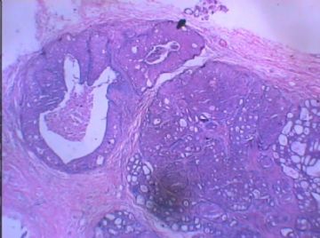
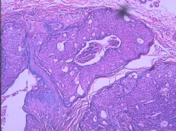
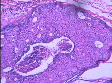
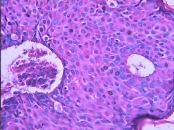
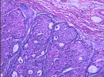
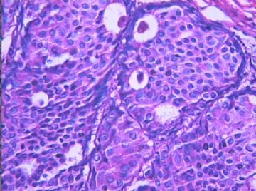
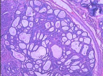
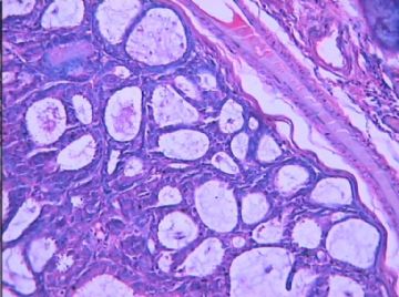
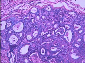
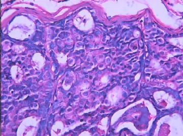
- B1572乳腺低级别导管内癌?
| 姓 名: | ××× | 性别: | 女 | 年龄: | 36岁 |
| 标本名称: | 左侧乳腺包块 | ||||
| 简要病史: | |||||
| 肉眼检查: | |||||
相关帖子
- • 左乳腺肿物
- • 乳腺癌?
- • 乳腺肿物
- • 乳腺肿物
- • 左乳癌标本乳头一个导管内的病变
- • 乳腺两个相邻导管内的病变
- • 乳腺肿物,请各位老师帮忙会诊
- • 女 46岁发现左乳腺肿块一月余
- • 乳腺肿物
- • 乳腺肿物
It is true that central necrosis can occur in low or high grade DCIS, atypical ductal hyperplasia, and usual UDH. I call DCIS with comedo necrosis only in the condition of DCIS with nuclear grade 3. In other words most people including oncologists, breast surgens think comedo DCIS is high grade lesion. We report nuclear grade 1 or 2 DCIS with necrosis as DCIS with central necrosis, nuclear grade 1 or 2, not mentioning comedo necrosis. Some people call nuclear grade 2 DCIS with necrosis as comedo necrosis also.
Agree with Dr. Shen that the last four photos alone may not be DCIS. However, the first six photos demostrate proliferation of relative monotonous, uniform, round cell popularion with mild variable nuclei in size ans shape. The round microlumens are present. I have no doubt that they are DCIS. They are not high grade (nuclear grade 3). I fell the nuclear grade 1.6. Ha, ha. In fact it is easy to reconganize if DCIS is nuclear grade 3 or grade 1. If you are not sure, just call nuclear grage 2. In this way you will be always right.
For DCIS you should report:
Nuclear grade: 1 (low grade), 2 (intermidiate), 3 (high grade)
Type or pattern: solid, cribriform, papillary micropapillary, comedo, clear cell, apocrine, basal-like et al.
Necrosis or calcification.
-
本帖最后由 于 2008-12-03 21:54:00 编辑
From 笃行者
诚然,坏死并不能鉴别UDH、ADH、DCIS.但认为在DCIS的分级中有意义。
我们所掌握的标准是:DCIS 核1级时---低级别DCIS
核2级时---中间级别DCIS
核3级时---高级别DCIS
但是,如果低级别(1级)的核,加上有粉刺样坏死,则诊断中级别DCIS
中间级别(2级)的核,加上粉刺样坏死,则诊断高级别DCIS
不知当否,请指教。
Thank Dr. 笃行者 for reading my comment. Sorry I even do not know how to call the first letter of your chosen name. My Chinese teacher did not teach me well. Ha, Ha.
There are many different classification of DCIS. I list few examples here
1. Scott (modified Lagios system)(Scott MA et al. Ductal carcinoma in situ of the breast: reproducibility of histological subtype analysis. Hum Pathol. 1997;28(8):967-973)
A. Low grade: Noncomedo histology, low nuclear grade and no necrosis.
B. Intermediate grade: Intermediate histology, intermediate nuclear grade and no necrosis.
C. High grade: Comedo histology, high nuclear grade and extensive necrosis.
2. European Pathologists Working Group: (Holland R. et al. Ductal carcinoma in situ: a proposal for a new classification. Semin Diagn Pathol. 1994;11(3):167-180.
A. well differentiated: Evenly spaced, markedly polarized nucleiof uniform size and regular comtour with chromation, inconspicuous nucleoli, and rare mitotic figures. Necrosis absent or minimal.
B. Intermediately differentiated: Mildly or miderately pleomorphic nuclei with some polarization and variation in size, comtour, and distribution. Fine to coarse nuclear chromatin and small nucleoli.
C. Poorly differentiated: Highly pleomorphic, poorly polarixed nuclei with irregular contour and distribution. Coarse, clumped chromation and prominent nucleoli. Central and individual necrosis often present.
abin译:
谢谢Dr.笃行者阅读我的评论。抱歉,我甚至不会读"笃"字,我的中文都教得不好,哈哈。
DCIS有很多不同分类,我列举一些:
1. Scott (modified Lagios system)(Scott MA et al. Ductal carcinoma in situ of the breast: reproducibility of histological subtype analysis. Hum Pathol. 1997;28(8):967-973)
A. 低级别(组织学无粉刺样坏死),低核级别,无坏死。
B. 中级别(组织学中间状态):中度核级别,无坏死。
C. 高级别(组织学粉刺样坏死):高核级别,广泛坏死。
2. European Pathologists Working Group: (Holland R. et al. Ductal carcinoma in situ: a proposal for a new classification. Semin Diagn Pathol. 1994;11(3):167-180.
A. 高分化:分布均匀,核极性明显,大小一致,轮廓规则,核仁不明显,罕见核分裂像。坏死无或轻微。
B. 中分化:轻到中度多形性核,存在一些极性,大小、轮廓和分布有差异。核染色质纤细到粗糙,有小核仁。
C. 低分化:高度多形性,核极性私愤,轮廓和分布不规则。粗糙、块状染色质,核仁明显。存在中央和单个细胞坏死。
-
本帖最后由 于 2008-12-03 22:20:00 编辑
Continue my writing:
3. Silverstein et al. Prognostic classification of breast of ductal carcinoma in situ. Cancer 1995:345:1154-1157. Silverstein proposed a classification of DCIS based on nuclear grade(high or nohigh) and the presence or absence of necrosis as part of a prognostic index.
No single grading system for intraductal carcinoma has been demonstrated to notably superior, and none has gained widespread acceptance. However, a pathology report for DCIS should provide information about the descriptive characteristics considered to be essential in most grading system. The three essential elements noted were nuclear grade, necrosis, and architectural pattern. So for DCIS, we just report nuclear grade , architectural (or growth) patterns (solid, cribriform, et al), necrosis and microcalcification if they are present. It is easy to call nucear grade 1 and 3 immediately. It is nuclear 2 if you are thinking what it will be. We report Nottingham grade for invasive carcinoma.
In term of your question, the grad should be increased if necrosis is present. I did not see the study report s about this and cannot evaluate it. It is not unusual to see UDH with central necrosis. I just wonder it can be called ADH. I do not think so.
I think the most importance for pathologists is that we and surgens or oncologists use the same lanuages. We should be consistent in our pathology reports. They know what we are talking about.
Thank you. I have to read some book and papers for your interesting question.
Hope some one can translate into Chinese.
abin译:
接上:
3. Silverstein(Silverstein et al. Prognostic classification of breast of ductal carcinoma in situ. Cancer 1995:345:1154-1157)根据核级别(高或非高级别)和是否存在坏死作为预后指数。
没有哪一个导管内癌的分级系统被证明具有明显的优越性,也没有任何一个得到广泛接收。然而,DCIS的病理报告中应当提供足够信息,包括大多数分级系统中认为重要的描述性特征。三个重要方面包括核级别、坏死和结构类型。因此,对于DCIS,我们只报告核级别、结构或生长模式(实性、筛状等)、坏死和微钙化如果存在也要报。核级别1和3很容易取,只是核级别2很费思考。我们用Nottingham分级报告浸润性癌。
对于你提到的,如果坏死存在,分级就上升。我没有见过这方面的研究报告,不好评价。UDH伴中央坏死也并不少见。我只会考虑它是否可能为ADH。我不这样认为。
我认为对病理学家最重要的是,我们和外科医生和肿瘤学家使用相同语言。我们应当在病理报告中一致起来。他们知道我们在讲什么。
谢谢你。你的问题,我要看书查资料才能回答。

