| 图片: | |
|---|---|
| 名称: | |
| 描述: | |
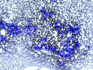
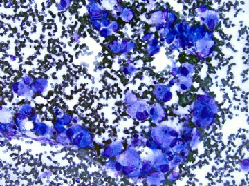
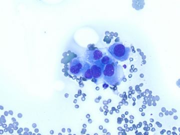
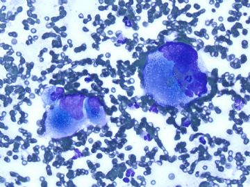
- Lung mass FNA today Please join in the discussion
Old man with a lung mass 3 cm.
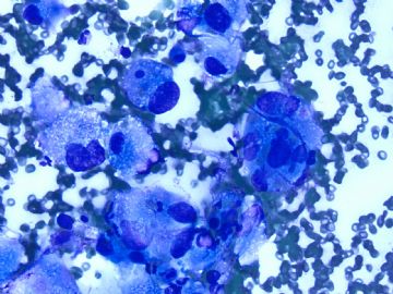
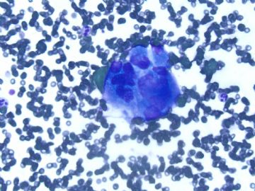
Radiologist did CT-guided FNA and I did on site evaluation this afternoon. I called malignant cells based on above one DQ. The procedure was stopped because patient had bleeding and also I think I should have enough cells for a cell block. Cytopathologists are required to give diagnosis (at least malignant, atypical, benign) on site in our institute if it is possible.
In fact I really do not know what kinds of tumor for this case. I have not seen the Pap stain yet. I have my differential dx and ordered some IHC already.
Hope people who see this case write down your differential dx and IHC.
When I have IHC results I will put here.
-
shn-821128 离线
- 帖子:277
- 粉蓝豆:3
- 经验:277
- 注册时间:2008-11-02
- 加关注 | 发消息
Thank Dr. 天山望月 and 月新' s great translation.
Conventional RCC=clear cell RCC, most common type of RCC: most cases are CK7 negative, occasional cases can be CK7 negative.
Papillary RCC: most of cases are CK7 positive as Dr. Chen has mentioned..
GU pathology=genitourinary pathology
GI pathology=gastrointestinal pathology
Sorry to cause the confusion.
-
本帖最后由 于 2008-12-10 22:14:00 编辑
非常感谢赵老师的认真和热情,中国病理医生可能不太熟悉这种染色,因此他们参加讨论不太积极,不太敢发言,大家内心学习的热情是非常高的,我也不习惯这种染色。但是赵老师的思路太好了,分析也特别到位,最后的病理报告写的更是句句珠矶,把病理报告书写的如此精致,把与患者住院医生的交流,此切片与某某某细胞病理专家会诊,与某某泌尿病理专家会诊,都记录在案。成为可能永久保存并且永远的证据,这真是太好了。而且病理报告留有充分的余地。不说过头话,这是真正的病理报告。值得我好好学习。
一个病例讨论,收益匪浅。有矛塞顿开之感。
一个字,好哇!
中国的病理医生,加油。
-
本帖最后由 于 2008-12-22 12:24:00 编辑
Thank Dr. Chen, 天山望月's discussion and 月新老师的翻译.
I put this topic here for 2 weeks already.我准备将本例放两周时间, I hope my colleagures in China can join the discussion. 希望中国病理医生同行们参加讨论。In fact it seems that I and dr chen are main persons in discussion here.事实上,似乎主要是由我和陈大夫参加了讨论。We do not need to put cases here if we want to discuss some cases.如果光我们自己讨论一些病例的话,并不需要在此讨论。 You need to be more active if you think you are weak and want to learn cytopathology.大家更需要积极参与,如果知道我们的不足,如果我们想提高,想学习细胞病理学的话,就一定要参加讨论。 Anyway I summarize the case now. 无论大家的积极性如何我把本例小结如下。 i sign out the case as following:我的病理报告是这样书写如下:
FINAL DIAGNOSIS:最后诊断: Lung Mass, left, CT-Guided Fine Needle Aspiration Biopsy"左肺包块CT导引下细针穿刺活检 SATISFACTORY FOR INTERPRETATION.获得细胞学质量满意。 POSITIVE FOR MALIGNANT CELLS.查见恶性细胞。
POORLY DIFFERENTIATED NON-SAMLL CELL CARCINOMA (SEE COMMENT).
COMMENT:低分化非小细胞癌(详细讨论如下)
The aspiration smears are cellular and reveal single and dyschhesive clusters of malignant cells in a blood background.本例穿刺细胞涂片见血性背景中有许多恶性细胞,细胞呈单个或散在团块状排列。 The tumor cells are large in size, very pleomorphic, and have abundant, vacuolated to dense cytoplasm, and hyperpchromatic nuclei with irregular contours and prominent nucleoli.瘤细胞个大,多形性明显,浆丰,胞浆从空泡状到致密,核深染,外形不规则,核仁明显。 Cell block contains similar cells with smears. 细胞块切片细胞类似涂片细胞。To further cahracterize the tumor cells, immunohistochemical studies were performed on the cell block sections with the following results. 为了进一步的研究本例,用细胞块做了相应的免疫组化。结果如下: Lists of all the IHC results (I have mentioned above)详细结果可以查免疫组化上一楼的帖子(我已经提供)。 Combined the cytomorphologic and immunophenotypic findings are of a poorly differentiated non-small cell carcinoma.结合细胞形态学和免疫组化表达,结果是低分化非小细胞癌。 The immunoprofile of this lesion is similar to the neck mass (case No) and this may represent similar or same tumor.本例的免疫组化表达类似该患者颈部包块,这可能是相同表达或者就是同一肿瘤。 No slides of previous case are available for review. 没有复习该患者前一次的切片,The differential diagnosis includes but is not limited to poorly differentiated adenocarcinoma of lung or metastatic carcinoma from nasopharyngeal origin, upper gastrointestinal origin including esophagus, stomach, pancreatobillary sites.鉴别诊断可以包括但不能局限于肺低分化腺癌、转移性鼻咽癌、上消化道包括食道、胃、胰腺部位的癌肿 In review of CA9, vimentin, and CD10 immunostain positivity in the present cells, it raises the possibility of metastatic renal cell carcinoma.复习CA9, vimentin, and CD10阳性表达,转移性肾癌可能性很大, Clincal correlation with imaging is recommedded.建议结合影象学和临床考虑。
I discussed the case with Dr. xxxx (primary physician) at 3:30 PM, on m/d/y. Dr. xxxxx (cytopathologist) and Dr. xxx (GU pathologist) have reviewed the case and concur with above interpretation.我与病人的主管医生XXX一齐讨论该病例在某年某月某日,下午 3:30 ,与细胞病理专家XXX以及泌尿病理专家XXX一齐复习了该病例。 Above is present in my full final report.上述是我们全部的最后报告
Share some thought with you guys:有一些想法与同道分享。 Previous surgical specimen is a neck mass diagnosed as poorly differentiated carcinoma.过去患者颈部包块手术标本诊断该病人为低分化癌, The origin of the neck mass was not known.恶性肿瘤的起源并不清楚 I do not think the review of these slide can help me and this why I did not ask for these slides.我认为过去的切片并不一定能帮助我们,所以我并没有复习过去的病理切片, This is complicated case and I cannot figure out the origin on FNA.这是一个复杂的病例,我用细针穿刺涂片也不能确定组织学起源。 It is fine that people know the limitation of the FNA cytology and even surgical pathology.非常幸运的是大家都知道细针穿刺的局限性,甚至手术切除标本病理学也有诊断的局限性, In fact I will choose metastatic RCC if this is test, but not a true case. 事实上如果这是一例考试病例,我可能选择是转移性肾细胞癌,但这不是考试。
Support RCC:支持转移性普通型肾细胞癌如下:
1.Multiple locations of metastasis
1、患者为多部位转移,
2.Cytologic features (even though the cytology of most RCC cases is not so ugly).
2、 细胞学特点,特别是许多普通型肾细胞癌的细胞学表现不是那么的怪异,
3. IHC results: positive for CD10, vimentin, CA9. CK7 is positive for papillary RCC.
3、免疫组化结果CD10, vimentin, CA9. CK7阳性,支持肾乳头状肾细胞癌。
CK7 is negative in most conventional RCC cases, but some conventional cases can be positive.多数普通型肾细胞癌CK7是阴性,有时是阳性, We cannot rule out RCC based on the positive CK7.根据CK7阳性不能排除肾细胞癌。 I feel unconfortable about RCC 不支持肾细胞癌如下: The clinician said no lesion in kindey in imagining result.临床和影像学不支持肾细胞癌, I do not understand what carcinomas can be EMA purely negative.我不理解为什么只有EMA是单纯阴性, Anyway this is what I can do for this case. 就本例来讲,我也只能做这些, Suggest to you:建议大家:
1. Know the limitation of cytopath and pathology.
1、要明白细胞病理和组织学病理学都有自己的局限性,
2. Do not need to make a definite dx if you are not sure. Leave some spce for you.
2、如果你不确定不要做明确的诊断,给自己留点余地。
3. Contact with the primary doctors to know the clinical information,
3、与患者的经管医生接触了解更多的临床知识,
let him know your interpretation.让临床医生知道你想要表达的意思, Record the comunication in your final report for some difficult case.某些疑难的病例,在你最后书写的病理报告中用文字记载下来这些交流内容,
4. Show your case with other pathologists for some difficult cases and record in your report.
4、把困难的病例拿出来与其它病理医生交流,并且记录在案。
Thank for reading and discusing the case.
感谢大家阅读并且参加讲讨论。
-
本帖最后由 于 2008-12-09 23:33:00 编辑
(续译)
和朋友们分享一些思考:
以前颈部肿块的手术标本诊断为低分化癌。颈部肿块的起源不明。我认为复阅以前的这些切片对本例没有帮助,这是为什么我没有要这些切片的原因。这是复杂的病例,在FNA上我无法确定肿瘤起源。所幸的是人们知道FNA的局限性和甚至外科病理也有局限性。事实上,如果考试,我将选择转移性的RCC(肾细胞癌),但真正病例不能这样。
支持RCC:
1.多发性转移
2.细胞学特点(尽管大多数RCC病例的细胞并非如此的恶)
3.免疫组化结果: CD10,vimentin, CA9阳性。 乳头状肾癌呈CK7阳性。 CK7在大多数乳头状RCC呈阴性,但某些普通型RCC可以是阳性。我们不能根据CK7 阳性来排除RCC。
关于RCC,我感觉unconfortable
临床医生说,肾脏的影像学检查没有病变。
我不明白什么癌EMA是完全的阴性。
反正对于这此例,我只能做到这样。

- 广州金域病理




 我学习中
我学习中


