| 图片: | |
|---|---|
| 名称: | |
| 描述: | |
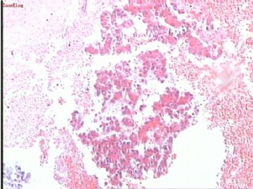
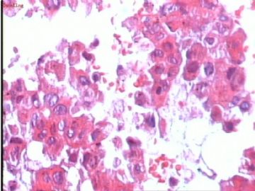
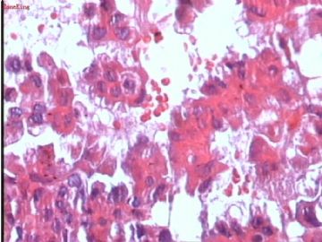
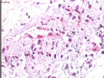
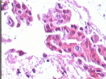
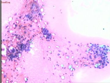
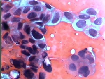
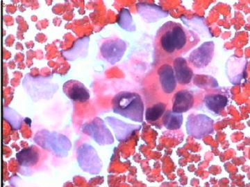
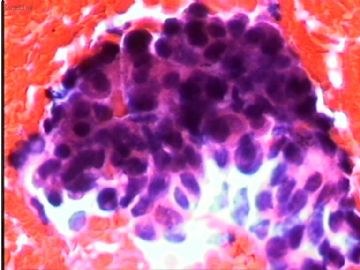
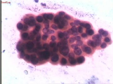
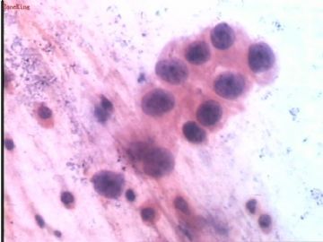
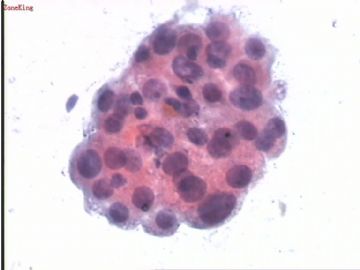
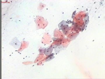
- 肺穿刺细胞,女,65岁。
-
In our hospital, we use H&E stain for cell block too. The Pap (巴氏染色) stain you showed in your pictures does not look like Pap stain. The hallmark of Pap stain is crisp and detailed nuclear morphology, which are missing in your stains. Maybe, you should work on the stains a little bit to improve.
-
13楼: If you did Pap stain on the FNA slides, even if you get a lot of blood, the stain still should be quite sharp. I suspect that people let the slides air-dryed, in another word, the slides did not get fixed by alcohol quickly. If the sample truly is very bloody, we also using liquid based cytology and process the specimen as a ThinPrep Pap slide, with the ThinPrep filter, we can get rid a lot of blood and junks. It is expensive though. Hope the above information help.
-
The quality of the slides are not very good. What stain is this? Is it quick H&E? If that is the case, I strongly discourage this type of stain. I basically agree with Dr. Zhao, this is non-small cell carcinoma. If this tumor is surgeically removable, I usually do not bother to do immunostains to further classify. If it is not resectable, because there is treatment differences between squamous cell ca and adenocarcinoma, I will do immunostains to help me further classify. This case I favor an adenocarcinoma on the cell block. One more comment, I have looked at some pictures people posted on this websites, the quality of the slides is not good, that is one of the main reasons to hinder the diagnosis.




 你的讨论非常有用!


