


| 图片: | |
|---|---|
| 名称: | |
| 描述: | |
- B1053女性,47岁,乳腺不明显的肿物切除.
| 姓 名: | ××× | 性别: | 年龄: | ||
| 标本名称: | |||||
| 简要病史: | |||||
| 肉眼检查: | |||||

名称:图1
描述:图1

名称:图2
描述:图2

名称:图3
描述:图3

名称:图4
描述:图4

名称:图5
描述:图5

名称:图6
描述:图6

名称:图7
描述:图7

名称:图8
描述:图8
标签:SPC 实性乳头状癌 EDCIS

- 病理,让疾病明明白白。
相关帖子
×参考诊断
非常感谢这楼上各位老师的分析,特别感谢Dr.Zhao和Dr.Stevenshen的深入讨论。
习惯查阅中文资料的朋友,可以参考《中华病理学杂志》2006年第10期上的两篇文献,谈到SPC和EDCIS以及它们之间的关系。我们论坛以前也有关于EDCIS的讨论
(乳腺冰冻之五--F55Y乳头溢液http://www.ipathology.org.cn/forum/forum_display.asp?classcode=148&keyno=30689&pageno=1)。
华夏病理/粉蓝医疗
为基层医院病理科提供全面解决方案,
努力让人人享有便捷准确可靠的病理诊断服务。
-
stevenshen 离线
- 帖子:343
- 粉蓝豆:2
- 经验:343
- 注册时间:2008-06-03
- 加关注 | 发消息
-
本帖最后由 于 2008-11-06 18:28:00 编辑
Many thanks to Dr. Zhao for the clear description and reference. I will pay more attention to solid papillary carcinoma and read the original article. All DCIS regardless of growth pattern are associated with risk of invasive carcinoma. The reason I prefer the diagnosis of solid DCIS, becaused it is also associated with cribriform DCIS or ADH nearby. Great case and I enjoy it.
abin译:
非常感谢Dr. Zhao详细讲述并提供文献。我会更加关注SPC并阅读原文。除了实性型DCIS,所有DCIS都有进展为浸润癌的风险。我倾向于诊断实性DCIS的原因,是因为它周围也伴随筛状型DCIS或ADH。病例非常好,我很喜欢。
-
本帖最后由 于 2008-11-06 18:24:00 编辑
For this case I have not any doubt this is a solid papillary carcinoma.
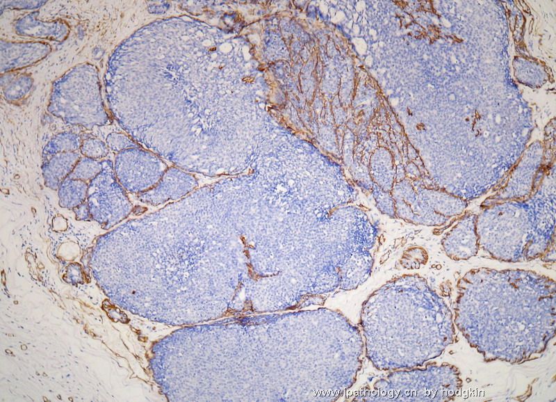
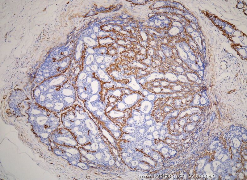
SPC defined by Maluf sndf Koerner (Solid papillary ca of the breast: a form of intraductal carcinom with endocrine differentiation frequently associated with mucinous carcinoma. Am J Surg Pathol. 1995;19:1237-1244). : These tumors had the characteristic appearance showing nodular proliferations of neoplastic cells in which each nodue appeared to represent a distendeed well-cricumscribed duct with a thickened fibrous wall. Broad bands of dense fibrotic stroma with no or little intervening breast tissue separated individual nodule. The cellular proliferation solid in architecture but supported in all cases by hyalinized fibrovascular cores. Often it is a diagnosis in low power. We can appreciate some fibrovascular cores in fig 1, 3, 4. above case. Myoepithelial cells in periphery can be present or absent in SPC.
DCIS with solid growth pattern and PSC are not the same terminology. PSC has its charateristic appearance as mentioned above.
Lack of myoepithelial cells around the periphery of the neoplastic nodules in some cases raise the possibility that some of SPCs may represent circumscribed nests of invasive ca rather than variant of DCIS.
In this case myoepthelial cell in the periphery are present. So it is just a variant of DCIS. Clinical prognosis is similar to DCIS.
Abin: I will appreciate if you can translate this part into Chinese. Thanks, cqzhao
abin译:
本例我毫无疑问地认为它是SPC。
1995年Maluf等提出SPC(Solid papillary ca of the breast: a form of intraductal carcinom with endocrine differentiation frequently associated with mucinous carcinoma. Am J Surg Pathol. 1995;19:1237-1244)。SPC特征性的表现为:肿瘤细胞结节状增生,每个结节呈界清的扩张导管结构,围绕厚的纤维壁。致密的纤维性间质,呈宽束状,其中有少量或无乳腺组织穿插,这些乳腺组织由单个结节分隔的。在所有病例,这些富于细胞的增生呈实性结构,但有透明变性的纤维血管轴心支持。这种特征通常以低倍镜下即可诊断。我们可以在图1、3、4中辨认一些纤维血管轴心。SPC中结节周围的肌上皮细胞可以存在也可能缺失。
实性生长型DCIS和SPC不是同义词。SPC有自己特征性的表现,如上所述。
部分病例中,肿瘤结节周围缺乏肌上皮细胞,这一现象提出一种可能性:部分SPC可能是界清的浸润性癌巢,而不是DCIS的亚型。
本例,结节周围存在肌上皮细胞。因此这例只是DCIS的一种变型。临床预后与DCIS相似。
(abin附:晚上要飞到青岛去,我得在离开前完成任务,呵呵。谢谢Dr.Zhao详细讲解)
-
stevenshen 离线
- 帖子:343
- 粉蓝豆:2
- 经验:343
- 注册时间:2008-06-03
- 加关注 | 发消息
-
本帖最后由 于 2008-11-06 17:58:00 编辑
It's very interesting that we look at the same picture but have different interpretation. I did not see obvious papillary lesion as Dr. Zhao. My interpretation of this lesion is: DCIS of solid growth pattern with cancerization of adjacent lobules. It is also associated with columnar cell hyperplasia and ADH. We all seem to agree it is DCIS and the risk of invasive cancer and treatment should be the same. Thanks.
abin译:
我们经常会遇到这种情况,看到相同图片却产生不同见解,这很有趣。我没有像Dr.Zhao看到明显的乳头状病变。我的观点是:实性生长型DCIS,伴邻近小叶癌化。也伴有柱状细胞增生和ADH。我们似乎都同意它是DCIS性质和进展为浸润癌的风险以及相同的临床处理。
谢谢。
-
本帖最后由 于 2008-11-05 20:31:00 编辑
See the photos again.
There are two diagnosis lines
1. Solid papillary carcinoma (Why? first four photos show large circumscribed celluar nodeles separated by fibrous tissue).
2. Atypical papilloma (other photos)
abin译:
再次看图。
列出两个诊断:
1.SPC(为什么?前四图显示大面积界清的富细胞性结节,由纤维组织分隔)。
2.不典型乳头状瘤(其它图)。
-
本帖最后由 于 2008-11-05 20:28:00 编辑
1. Solid papillary carcinoma without frank invasion, variant of DCIS.
2. Last four photos: atypical papilloma vs focal DCIS within Papilloma (it is not important because you have above diagnosis already)
abin译:
1.无明显浸润的SPC,DCIS的亚型。
2.后四图:不典型乳头状瘤/乳头状瘤内局灶性DCIS(这一条不太重要因为已经有了上述诊断)。
-
stevenshen 离线
- 帖子:343
- 粉蓝豆:2
- 经验:343
- 注册时间:2008-06-03
- 加关注 | 发消息
-
本帖最后由 于 2008-11-05 20:22:00 编辑
First four photos seem to be solid papillary carcinoma. Suggest IHC for myoepithelial markers.
Solid papillary carcinoma: absence of myoepithelial cells within the cellular proliferation is chracteristic. Myoepithelial cells around the peripheries of neoplastic nodules may be absent also. The tumor can have endocrin features with positive stains for chromogranin and synaptophysin. It is a variant of DCIS. Some breast pathologists think that at least some of the cases may represent nests of invasive carcinoma (if no myoepithelial cells around the peripheries). Anyway solid papillary carcinoma has an indolent clinical course. Just for your reference.
abin译:
前四图似乎是实体性乳头状癌(SPC),建议检测肌上皮标记。
SPC:在细胞丰富的增生区域缺乏肌上皮,具有特征性。肿瘤性结节的周围,肌上皮细胞也可能缺失。肿瘤可有神经内分泌特征,嗜铬素(CgA)和突触素(SYN)阳性。它是DCIS的一种亚型。一些乳腺病理学家认为至少部分病例可以出现浸润癌巢(如果周围缺少肌上皮细胞)。总之,SPC在临床上呈惰性过程。
仅供参考。
-
liguoxia71 离线

- 帖子:4174
- 粉蓝豆:3122
- 经验:4677
- 注册时间:2007-04-01
- 加关注 | 发消息


