


| 图片: | |
|---|---|
| 名称: | |
| 描述: | |
- 颈4椎体肿瘤
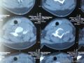 图1
图1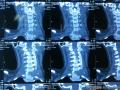 图2
图2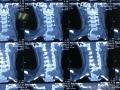 图3
图3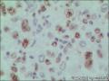 图4
图4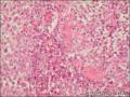 图5
图5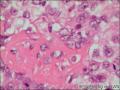 图6
图6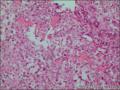 图7
图7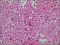 图8
图8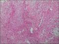 图9
图9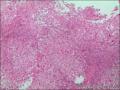 图10
图10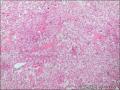 图11
图11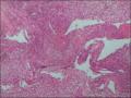 图12
图12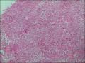 图13
图13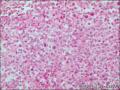 图14
图14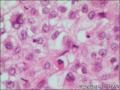 图15
图15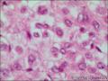 图16
图16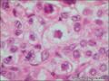 图17
图17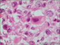 图18
图18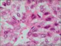 图19
图19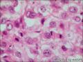 图20
图20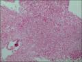 图21
图21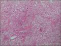 图22
图22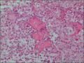 图23
图23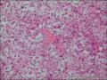 图24
图24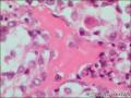 图25
图25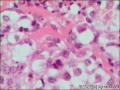 图26
图26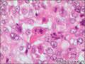 图27
图27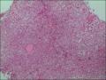 图28
图28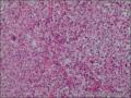 图29
图29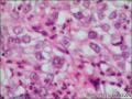 图30
图30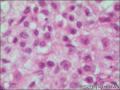 图31
图31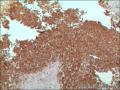 图32
图32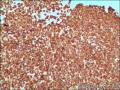 图33
图33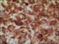 图34
图34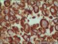 图35
图35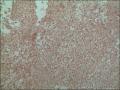 图36
图36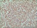 图37
图37
| 性别 | 女 | 年龄 | 20岁 | 临床诊断 | 颈4椎体肿瘤 |
|---|---|---|---|---|---|
| 一般病史 | 颈部疼痛不适3月 | ||||
| 标本名称 | 颈4椎体肿瘤 | ||||
| 大体所见 | 灰白碎组织,大小1.6*1.5*0.8cm | ||||
标签:肿瘤
相关帖子
×参考诊断
引用 5 楼 chz1970 在 2013-10-03 00:03:59 的发言:
王老师,您好!在IHC染色结果出来前,我们的思路和您一样,但IHC染色结果让我们很困惑!AE1/AE3 3+,CAM5.2 3+,EMA 3+,Vim3+,Calponin 1+,CD99 灶状弱阳性,PET-CT及病史、体检均未见其他部位有肿瘤存在!组织学像骨肉瘤,IHC提示为转移性癌,所以向各位老师请教!
尤文肉瘤/PNET是可以有上皮分化而表达上皮性标志物的。
我们知道,小细胞骨肉瘤也有呈上皮样型的亚型,可以有菊形团结构,既可以表达NE标志物,也可提示表达上皮性标志物的。
让我们来看一看国外文献报道。

- 王军臣
-
本帖最后由 海上明月 于 2013-10-03 15:19:17 编辑
尤文肉瘤/PNET可表达上皮标志物角蛋白,在电镜下具体上皮性细胞的一些特征。
Int
J Surg Pathol. 2005 Jan;13(1):43-50.
Keratin-positive Ewing's sarcoma: an ultrastructural study of 12 cases.
Srivastava A, Rosenberg AE, Selig M, Rubin BP, Nielsen GP.
Source
Department of Pathology, Tufts-New
England Medical Center, Boston, MA 02114, USA.
Abstract
Ewing's sarcoma/primitive neuroectodermal tumor
(EWS/PNET) is an aggressive neoplasm of bone and soft tissue. Histologically,
it is characterized by the presence of small round blue cells, which usually
express MIC-2 and FLI-1 immunohistochemically. The most specific feature for
diagnosis, however, is cytogenetic or molecular evidence of a consistent
abnormality, the t(11;22)(q24;q12), or variants thereof. The immunohistochemical
expression of keratins in a significant proportion of these cases has been
highlighted in several recent studies. The ultrastructural features of these
keratin-positive tumors have not, however, been characterized in
detail. In this study we analyzed the ultrastructural features of 12
well-documented EWS/PNETs that stained strongly for pankeratin by
immunohistochemistry. Ultrastructurally, the tumor cells contained a few
organelles, which included a small number of mitochondria, poorly developed
Golgi complexes, free ribosomes, and inconspicuous rough-endoplasmic reticulum.
Rudimentary cell junctions were seen in 2 tumors while prominent
junctions were observed in the remaining 10. Fivetumors contained
intracytoplasmic filaments, and definite tonofibrils were identified in 2.
Well-developed basal lamina around tumor cells were also demonstrated in 2 tumors.
Follow-up information was available for all cases. Seven patients died of
disease, 2 are alive with disease, and 3 have no current evidence of disease.
The cohort includes 5 patients with a type-1 translocation, which has been
associated with a better prognosis in some studies; 4 of these patients have
died of their disease, and 1 is alive with recurrent disease. This study shows
that keratin-positive EWS/PNETs have evidence of epithelial differentiation
ultrastructurally, and may possibly represent a more aggressive subset of the
EWS/PNET group of tumors.
- 王军臣
文献报道的病例与本例肿瘤发生部位几乎相同,也是在颈椎脊髓脊膜部位发生的外周PNET/尤文肉瘤,表达CK阳性。
主要是要和其他类型小蓝圆细胞肿瘤相鉴别。
Int J Surg Pathol. 2005 Jan;13(1):93-7.
Cytokeratin-positive
meningeal peripheral PNET/Ewing's sarcoma of the cervical spinal cord:
diagnostic value of genetic analysis.
Woestenborghs H, Debiec-Rychter M, Renard M, Demaerel P, Van Calenbergh F, Van Gool S, Sciot R.
Source
Department of Pathology, University Hospital, Catholic
University of Leuven, Leuven, Belgium.
Abstract
Peripheral primitive neuroectodermal
tumor (PNET)/Ewing's sarcoma (ES) of the central nervous system is extremely
rare and should be differentiated from central PNET and other small blue
round cell tumors. We describe a case of a meningeal peripheral
PNET/ES of the spinal cord in an 11-year-old boy. Immunohistochemically, the small blue
roundcell tumor showed expression of epithelial markers and
of CD99, thus posing an important differential diagnostic problem with a poorly
differentiated synovial sarcoma. Fluorescence in situ hybridization revealed
rearrangement of the EWS gene, as seen in peripheral PNET/ES. Peripheral
PNET/ES does occur in the central nervous system, but its diagnosis can be
extremely difficult on morphologic and immunohistochemical grounds alone.
Genetic analysis plays a key role in its distinction from other small blue
round cell tumors.
- 王军臣