| 图片: | |
|---|---|
| 名称: | |
| 描述: | |
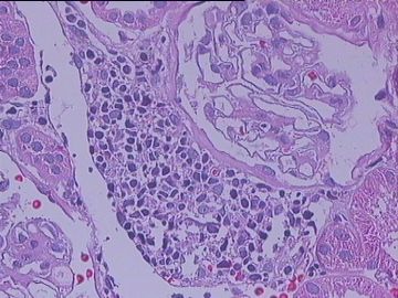
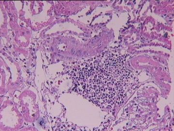
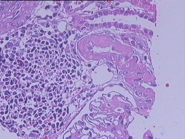
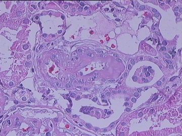
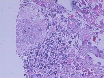
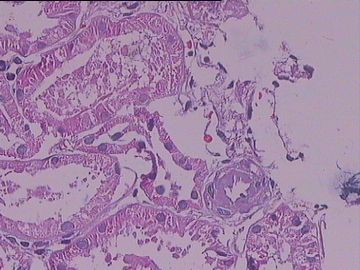
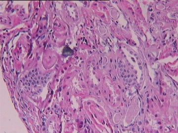
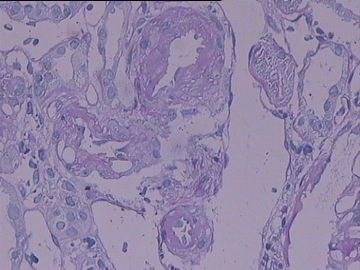
- 移植肾病理穿刺20101217
-
本帖最后由 于 2010-12-29 23:09:00 编辑
frankbj, thank you for posting this interesting case.
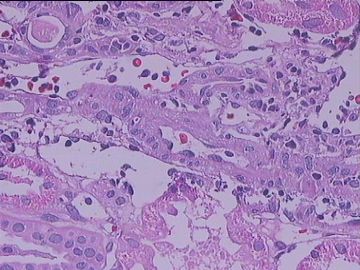
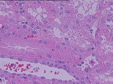
Once BK virus is identified, it is BK virus nephropathy, as seen in the above case. Often I was asked by nephrologist if there is co-existing cellular rejection. Some pathologists suggest if the tubulitis is 2 high power fields away from the BK virus infected tubules, the tubulitis should be considered as rejection-induced tubulitis, not infection-induced tubulitis. What is your approach to this issue?
I only started phenotyping the infiltrating mononuclear cells recently. I also heard if the infiltrating mononuclear cells are B-cell dominant, it is most likely BK virus nephropathy (of course, BK virus must be present). Let me know your thought about this.
-
本帖最后由 于 2011-01-04 23:22:00 编辑
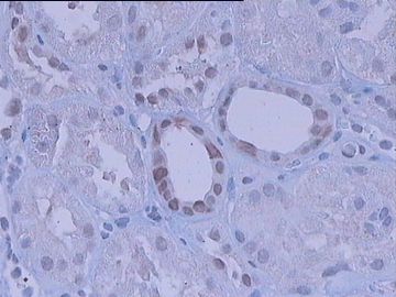
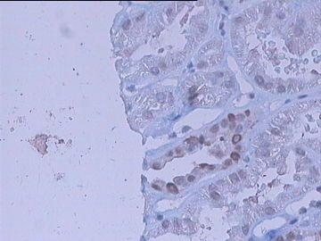
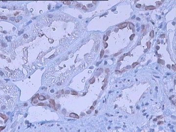
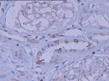
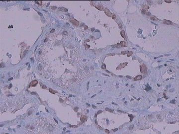
The above SV40 stains appear positive in the nuclei of tubular epithelium. But the pattern is slightly different from mine. The above positive stain is more in nuclear membrane. My positive stain often occupies the entire nucleus (please see the below photo).
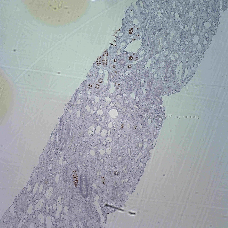
名称:图1
描述:图1
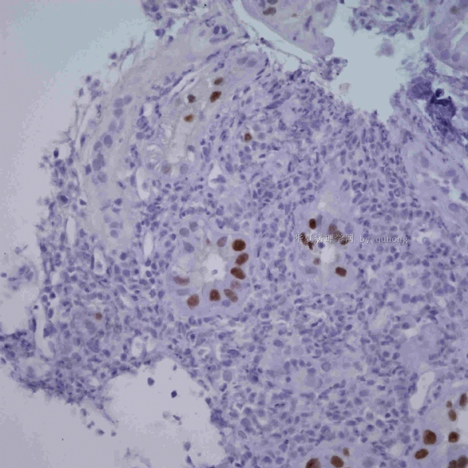
名称:图2
描述:图2
Here is the paper suggesting p53 stain+SV40.
Ann Clin Lab Sci. 2010 Fall;40(4):324-9.
Adjuvant role of p53 immunostaining in detecting BK viral infection in renal allograft biopsies.
Wiesend WN, Parasuraman R, Li W, Farinola MA, Rooney MT, Hick SK, Samarapungavan D, Cohn SR, Reddy GH, Rocher LL, Dumler F, Lin F, Zhang PL.
Dept. of Anatomic Pathology, William Beaumont Hospital, Royal Oak, MI 48073-6769, USA.
Abstract
BK virus infection is a significant threat to renal transplant outcome. Detecting viral infection in renal transplant biopsies using SV40 staining is less than ideal. SV40 antibody reacts with the large T-antigen of BK virus only at the early phases of infection and can miss cells in later stages of infection. As p53 is upregulated during both early and late phases of infection, this study set out to determine whether p53 staining could improve detection of BK virus infection in renal transplant patients. The control group consisted of 16 renal allograft biopsies without histologic evidence of BK virus infection, while the BK group consisted of 15 renal allograft biopsies with histologic evidence of BK virus infection. The biopsies from both groups were immunohistochemically stained with both SV40 and p53 antibodies. Dual staining with both markers was also performed to identify their nuclear co-localization. In the BK group, the percent of p53 staining (16.6 ± 4.8 %) was significantly higher than the percent of SV40 staining (5.4 ± 2.7%). BK virus infected cells revealed a unique p53 immunostaining pattern (strong nuclear staining with a central halo). Co-localization of SV40 and p53 was identified in cells that had characteristic nuclear features of BK virus infection by histology. The sensitivity and specificity for using p53 staining to identify BK infected cells was 92% and 86 %, respectively. In conclusion, p53 staining detects a higher percentage of BK virus infected cells than SV40 staining alone. Thus, for diagnosis of BK virus infection in renal allograft biopsies, p53 staining is a sensitive and specific method when used along with SV40 staining.

