共1页/9条首页上一页1下一页尾页
回复:9 阅读:1788
脑肿瘤?脓肿?这些增生的是血管母细胞吗?请会诊

LQING 离线

帖子:22
粉蓝豆:1
经验:42
注册时间:2008-01-02
加关注  |  发消息
楼主 发表于 2008-10-01 09:03|举报|关注(0)
浏览排序[ 顺序 逆序 楼主 支持 精彩 ]  快捷回复
姓    名: ××× 性别:   年龄:  
标本名称:  
简要病史:  
肉眼检查:  


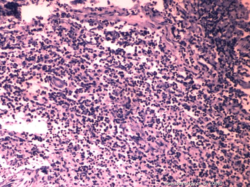
 男,42岁。反复头痛1月余。手术见颅内包块相临右颞筋膜增厚。切除包块及增厚筋膜。

软组织9×6×2.5cm,淡褐色,不规则,碎。

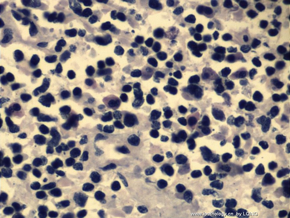
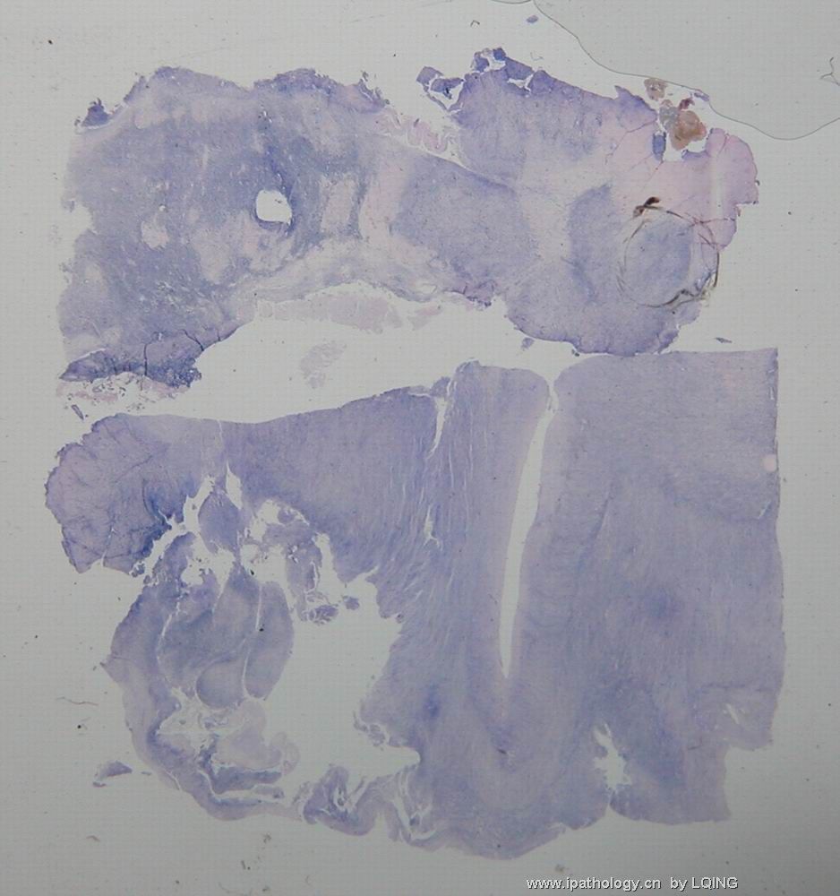
显微镜检查病灶内淋巴细胞浸润为主,伴酸性细胞浸润,部分区域血管增生,炎细胞浸润累及” 右颞筋膜”。拟诊断脑慢性脓肿。但切片免疫组织化学染色见密集ki-67阳性区。以下为免疫组织化学及ki-67阳性区的H.E。

  • 脑肿瘤?脓肿?这些增生的是血管母细胞吗?请会诊图1
    图1
  • 脑肿瘤?脓肿?这些增生的是血管母细胞吗?请会诊图2
    图2
  • 脑肿瘤?脓肿?这些增生的是血管母细胞吗?请会诊图3
    图3
  • 脑肿瘤?脓肿?这些增生的是血管母细胞吗?请会诊图4
    图4
  • 脑肿瘤?脓肿?这些增生的是血管母细胞吗?请会诊图5
    图5
  • 脑肿瘤?脓肿?这些增生的是血管母细胞吗?请会诊图6
    图6
  • 脑肿瘤?脓肿?这些增生的是血管母细胞吗?请会诊图7
    图7
  • 脑肿瘤?脓肿?这些增生的是血管母细胞吗?请会诊图8
    图8
  • 脑肿瘤?脓肿?这些增生的是血管母细胞吗?请会诊图9
    图9
  • 脑肿瘤?脓肿?这些增生的是血管母细胞吗?请会诊图10
    图10
  • 脑肿瘤?脓肿?这些增生的是血管母细胞吗?请会诊图11
    图11
  • 脑肿瘤?脓肿?这些增生的是血管母细胞吗?请会诊图12
    图12
  • 脑肿瘤?脓肿?这些增生的是血管母细胞吗?请会诊图13
    图13
  • 脑肿瘤?脓肿?这些增生的是血管母细胞吗?请会诊图14
    图14
  • 脑肿瘤?脓肿?这些增生的是血管母细胞吗?请会诊图15
    图15
标签:
0
添加参考诊断
×参考诊断
  

fyshan 离线

帖子:910
粉蓝豆:0
经验:968
注册时间:2006-10-04
加关注  |  发消息
1 楼    发表于2008-10-01 10:24:00举报|引用
返回顶部 | 快捷回复
 The surrounding area of the brain abscess usually show large amount of fibroblastic reaction, granulation reaction with increased vascular structure,  and gliosis, which may give an increased Ki-67 stain, this is a trick sign, don't misdiagnosed as tumor, Brain abscess has classic MRI presentation and pathology diagnosis can be confirmed by reticulin or trichrome stain to show the fibrosis (normal brain has no fibrosis, neither for brain tumor, unless this tumor is gliosarcoma), hopefully, this may help your dx. If you can submit a culture that will help your final dx.
0
回复

fyshan 离线

帖子:910
粉蓝豆:0
经验:968
注册时间:2006-10-04
加关注  |  发消息
2 楼    发表于2008-10-01 10:26:00举报|引用
返回顶部 | 快捷回复

 PS:

Please make sure those small blue cells are lymphocytes by immunostain for both T and B markers.

Frank

0
回复

lqing 离线

帖子:22
粉蓝豆:1
经验:42
注册时间:2008-01-02
加关注  |  发消息
3 楼    发表于2008-10-01 10:40:00举报|引用
返回顶部 | 快捷回复
以下是引用fyshan在2008-10-1 10:24:00的发言:

 The surrounding area of the brain abscess usually show large amount of fibroblastic reaction, granulation reaction with increased vascular structure,  and gliosis, which may give an increased Ki-67 stain, this is a trick sign, don't misdiagnosed as tumor, Brain abscess has classic MRI presentation and pathology diagnosis can be confirmed by reticulin or trichrome stain to show the fibrosis (normal brain has no fibrosis, neither for brain tumor, unless this tumor is gliosarcoma), hopefully, this may help your dx. If you can submit a culture that will help your final dx.

非常感谢.补充各图片名称:

图1,切片的cd20,图2,lca,图3syn.图4s-100.图5,6,7,8ki-67.图9ki-67阳性区的cd20.图10ki-67阳性区的vin.图11-14ki-67阳性区的HE.

0
回复

lqing 离线

帖子:22
粉蓝豆:1
经验:42
注册时间:2008-01-02
加关注  |  发消息
4 楼    发表于2008-10-02 10:54:00举报|引用
返回顶部 | 快捷回复
再上图.切片照片,显示淋巴细胞浸润不均.显微照片显示浸润细胞杂,有明显嗜酸性粒细胞.
  • 图1
  • 图2
  • 图3
0
回复

yourself 离线

帖子:663
粉蓝豆:7129
经验:691
注册时间:2007-06-30
加关注  |  发消息
5 楼    发表于2008-10-07 07:19:00举报|引用
返回顶部 | 快捷回复
 如确实如lqing所言,浸润的细胞为淋巴细胞、浆细胞,而且细胞均较成熟,可考虑脑脓肿(ki67可以高),但需排除淋巴瘤或淋巴瘤样肉芽肿,T和B标记看看结果如何?
0
回复

刻苦学习 离线

帖子:116
粉蓝豆:6
经验:122
注册时间:2008-12-22
加关注  |  发消息
6 楼    发表于2009-01-06 11:02:00举报|引用
返回顶部 | 快捷回复
 ?
0
回复

xiaohl 离线

帖子:282
粉蓝豆:24
经验:288
注册时间:2006-10-19
加关注  |  发消息
7 楼    发表于2009-01-07 20:46:00举报|引用
返回顶部 | 快捷回复

 考虑恶性淋巴瘤。

弥漫大B细胞淋巴瘤或淋巴瘤样肉芽肿。

需进一步标记 CD3,Kappa,Lamda,CD30,EBER原位杂交

0
回复

owen 离线

帖子:19
粉蓝豆:1
经验:19
注册时间:2009-01-02
加关注  |  发消息
8 楼    发表于2009-01-07 22:12:00举报|引用
返回顶部 | 快捷回复
 考虑恶性淋巴瘤
0
回复

mjma 离线

帖子:703
粉蓝豆:24
经验:789
注册时间:2006-09-28
加关注  |  发消息
9 楼    发表于2009-02-23 01:59:00举报|引用
返回顶部 | 快捷回复
I believe the lesion is most likely a large B cell lymphoma. 
0
回复
signature

聞道有先後,術業有專攻

回复:9 阅读:1788
共1页/9条首页上一页1下一页尾页
【免责声明】讨论内容仅作学术交流之用,不作为诊疗依据,由此而引起的法律问题作者及本站不承担任何责任。
快速回复
进入高级回复
您最多可输入10000个汉字,按 "Ctrl" + "Enter" 直接发送
搜索回复/乘电梯 ×
按内容
按会员
乘电梯
合作伙伴
友情链接